Targeted Molecular MRI of Colorectal Cancer by Antibody Functionalized Hyperpolarized Silicon Particles
Targeted Molecular MRI of Colorectal Cancer by Antibody Functionalized Hyperpolarized Silicon Particles
Whiting, N.; Hu, J.; Pudakalakatti, S.; McCowan, C.; Ramezani, S.; Davis, J.; Millward, N. Z.; Engel, B.; Liu, J.; Gellci, K.; Seo, H.; Brown, D.; Enriquez, J. S.; Menter, D. G.; Millward, S. W.; Gammon, S. T.; Piwnica-Worms, D.; Farach-Carson, M. C.; Carson, D.; Constantinou, P. E.; Bhattacharya, P.
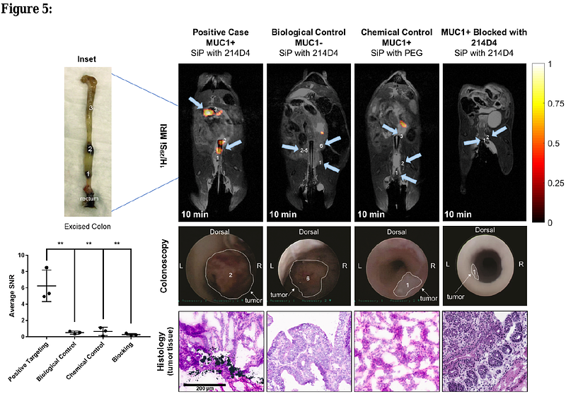
AbstractThe development of non-invasive, non-ionizing sensitive molecular targeting approaches to detect colorectal cancer (CRC) lesions is warranted to improve high risk patient outcomes. Hyperpolarized silicon nanoparticles and microparticles are potentially well-suited to act as targeted molecular imaging agents because of their overall biocompatibility and long-lasting enhanced magnetic resonance imaging (MRI) signals. In this study, dynamic nuclear polarization was performed on silicon particles functionalized with an antibody to Mucin-1 (MUC1), a surface mucin glycoprotein aberrantly expressed in CRC. Antibody conjugation to the particle surface did not affect 29Si hyperpolarization characteristics. Similarly, conjugation and the dynamic nuclear polarization process did not adversely affect the affinity of the targeting antibody. In vivo MRI scans performed 10-15 minutes after luminal administration of targeted hyperpolarized particles into human MUC1-expressing orthotopic CRC mouse models showed that particles actively targeted tumor sites. These results were supported by chemical and biological controls and blocking experiments as well as correlative immunohistochemical analysis. These surface-functionalized silicon particles are under development as a platform technology that will allow non-invasive molecular targeting of CRC using hyperpolarized MRI.