Deep Visual Proteomics links vascular smooth muscle cell phenotypes to atherosclerotic plaque stability

Deep Visual Proteomics links vascular smooth muscle cell phenotypes to atherosclerotic plaque stability
Kratz, E.; Sinha, A.; Adeshara, T.; Paloschi, V.; Sachs, N.; Pauli, J.; Glukha, N.; Huber, A.; Schanda, S.; Metousis, A.; Heymann, T.; Branzan, D.; Bleckwehl, T.; Hayat, S.; Maegdefessel, L.; Mann, M.
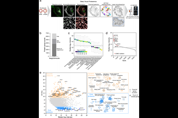
AbstractVascular smooth muscle cells (VSMCs) drive atherosclerosis through phenotypic switching, yet their spatial organization and protein signatures within plaques remain poorly characterized. Here, we applied Deep Visual Proteomics (DVP) to dissect more than 500 VSMC neighborhoods across 24 human carotid plaques and profile VSMC plasticity in disease. To functionally interpret these tissue proteomes, we built a reference atlas of primary VSMCs driven toward five phenotypes by TGF-{beta}, PDGF-BB, osteogenic stimuli, IL-1{beta}, or cholesterol, quantifying over 10,000 proteins. We integrated tissue and reference proteomes with a deep-learning framework that assigns functional phenotypes to each neighborhood. This revealed spatially distinct phenotype distributions and a shift toward dedifferentiated states in unstable plaques. Knockdown of four candidates (TNC, TNFAIP2, AEBP1, PLK1) validated operational roles during phenotypic switching. Our approach functionally annotates spatial proteomes and links VSMC plasticity to plaque instability in carotid artery disease.