Detection of vasculogenic mimicry in equine ocular, oronasal, and genital squamous cell carcinoma
Detection of vasculogenic mimicry in equine ocular, oronasal, and genital squamous cell carcinoma
Schwarz, S.; Kummer, S.; Klang, A.; Walter, I.; Nell, B.; Brandt, S.
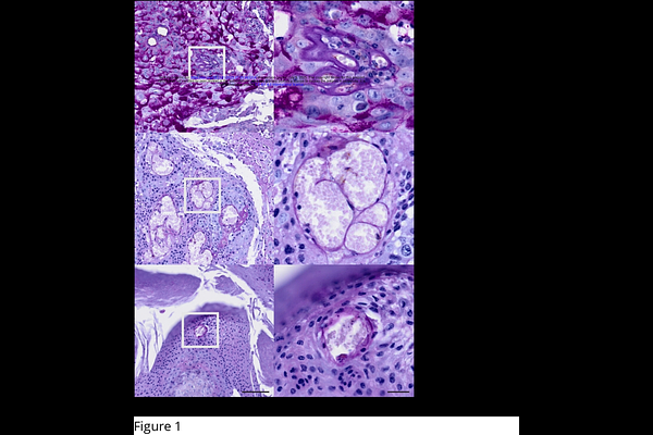
AbstractSquamous cell carcinoma (SCC) is the most common malignant tumor disease in horses. It predominantly affects the ocular, oronasal, and anogenital region. Equine SCC is difficult to treat, also because important aspects of SCC development and metastasis are still unclear. We previously provided evidence that equine SCC cells can adopt a stem cell-like phenotype as a hallmark of malignant progression. Here, we investigated whether equine SCCs harbor endothelial-like tumor cells that form an alternative network of pseudo-vessels better known as vasculogenic mimicry (VM). Following histopathological diagnosis, 43 equine SCCs or precursor lesions (15 ocular, 14 genital, 14 oronasal tumors) were PCR-screened for equine papillomavirus (EcPV) infection. Subsequently, formalin-fixed paraffin-embedded-sections of all tumors were analyzed by Periodic Acid-Schiff (PAS) reaction and immunohistochemical (IHC) staining for endothelial cell marker CD31. Obtained micrographs were evaluated by a scientific board to unanimously identify sections of intact tumor tissue displaying PAS-positive, CD31-negative lumens harboring erythrocytes. Thirteen lesions exhibiting these features were subjected to triple immunofluorescence (IF) staining for CD31, pan-cytokeratin (KRT) and type 4 collagen (Col4) or alpha smooth muscle actin (SMA) to confirm the presence of VM, and to determine whether pericytes have a role in this phenomenon. All genital and 50% of oronasal lesions scored positive for EcPV type 2, whilst ocular lesions tested negative. One mandibular SCC harbored EcPV type 5. Six genital, three oronasal, and four ocular tumors unambiguously exhibited VM as revealed by CD31-/PAS+ vessel-like structures containing erythrocytes, the detection of CD31-negative cells lining their lumens, and the presence of Col4 and SMA in this lining. Detection of these two proteins in the context of VM suggests that VM-forming cancer cells recruit pericytes to enhance channel formation and stability. To our knowledge, this is the first report providing evidence of VM in equine cancer, and more generally, SCC in animals.