Female Alms1-deficient mice develop echocardiographic features of adult but not infantile Alström Syndrome cardiomyopathy
Female Alms1-deficient mice develop echocardiographic features of adult but not infantile Alström Syndrome cardiomyopathy
McKay, E. J.; Luijten, I.; Thomson, A.; Weng, X.; Gehmlich, K.; Gray, G. A.; Semple, R. K.
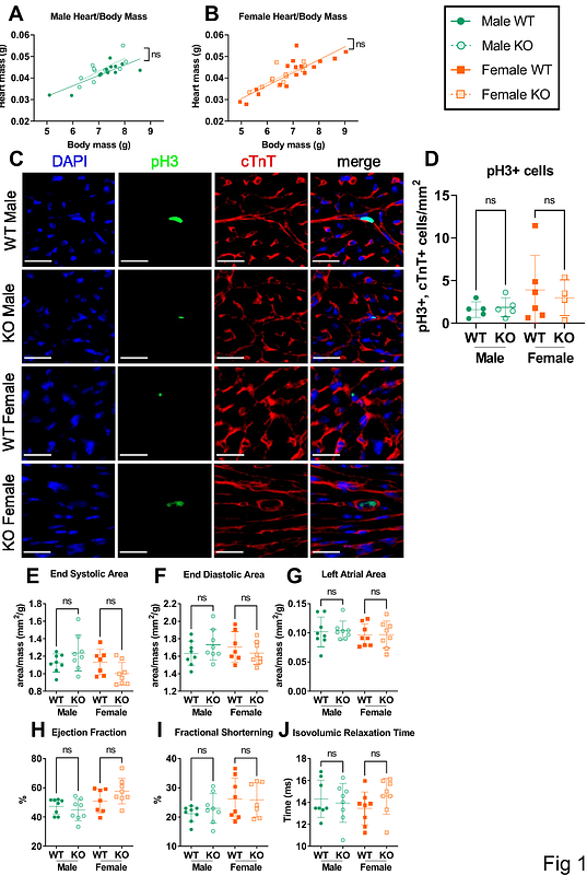
AbstractBackground: Alstrom Syndrome (AS), a multisystem disorder caused by biallelic ALMS1 mutations, features major cardiac complications often causing early mortality. These are biphasic, including infantile dilated cardiomyopathy, and distinct adult-onset cardiomyopathy. Cardiomyocyte maturation defects, cardiac fibrosis and early atherosclerosis have all been invoked as contributors to heart failure in AS, but their relative importance and inter-relationships are unknown. Methods: Cardiac function of global Alms1 knockout mice was assessed by echocardiography at postnatal day 15 (P15) and at 8 and 23 weeks of age. Echocardiography was also undertaken in female mice with Pdgfr-Cre-driven Alms1 deletion in cardiac fibroblasts and a small proportion of cardiomyocytes. Histological and transcriptional analysis of myocardium at P15 and 24 weeks of age was also performed. Results: Cardiac function was unaltered in knockout mice of both sexes at P15 and 8 weeks of age. At 23 weeks of age female but not male knockout mice showed increased left atrial area, decreased isovolumic relaxation time, and reduced ejection fraction, consistent with early restrictive cardiomyopathy. No histological or transcriptional changes could be identified in myocardium of 23-week old female Alms1 KO mice, however. Pdgfr-Cre-driven Alms1 KO in females did not recapitulate the phenotype of global KO at 23 weeks. Conclusions: Adult female, but not male, Alms1-deficient mice show echocardiographic evidence of cardiac dysfunction, consistent with the restrictive cardiomyopathy of AS. The explanation for sexual dimorphism remains unclear, but may involve metabolic or endocrine differences between sexes. No infantile cardiomyopathy was found in this study.