Observation of Field Cancerization in Human Breast Tissue with Raman Hyperspectral Imaging and Artificial Neural Networks
Observation of Field Cancerization in Human Breast Tissue with Raman Hyperspectral Imaging and Artificial Neural Networks
TAYLOR, J. N.; Bando, K.; Naoi, Y.; Fujita, S.
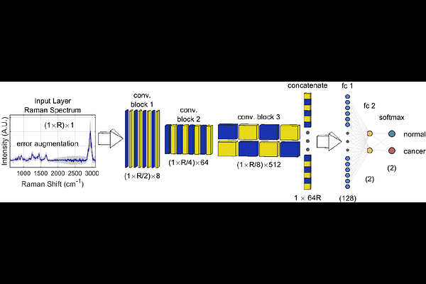
AbstractTissue-conserving partial mastectomy surgeries are often used to treat breast cancer, in which malignancies and surrounding margin tissues are removed. Morphologies of margin tissues are often assessed intraoperatively by a histopathologist, requiring excision and time-consuming preparation for microscopic examination. Changes in morphology often appear only after the mutation of cells involved in tumor propagation, suggesting that margins and perhaps conserved tissue may already contain cancer-converted cells prior to morphological changes. In a process known as field cancerization (FC), benign tissue surrounding a malignancy is affected by continued exposure to carcinogens, triggering cancer-promoting alterations like conversions of normal adipocytes and fibroblasts to cancer-associated adipocytes and fibroblasts. We report successful FC detection in human breast tissue samples excised during breast-conserving surgeries in human patients using Raman hyperspectral imaging and artificial neural networks (ANNs). Spectra in the images are segmented based on tissue modalities such as adipose, connective, and glandular tissues using intensities at Raman shifts of known molecules and label propagation, a method of transductive learning. ANNs that discriminate cancer from noncancer spectra return high quality (>95% accuracy) and reproducible distinction of spectra originating from cancerous tissue at high signal-to-error ratios (S/Es). Good distinction is returned in all modalities, with adipose-rich modalities returning the most reliable classifications, suggesting reliable discrimination of normal and cancer-associated adipocytes. Spectral S/Es are increased using larger spectral measurement areas, but this causes spectral mixing across different tissue modalities and significant biological variation, failing to deliver a corresponding increase in classification quality.