SPEND-hSRS imaging of fumarate uncovers mitochondrial metabolic heterogeneity
SPEND-hSRS imaging of fumarate uncovers mitochondrial metabolic heterogeneity
Sun, D.; Ding, G.; Lin, H.; Chen, G.; Wang, C.-C.; Bachoo, S.; Bohndiek, S. E.; Cheng, J.-X.
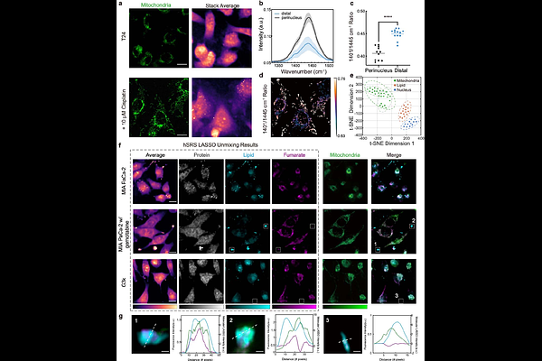
AbstractMitochondria, acting as the energy powerhouse, biosynthetic center, and reductive equivalent hub of the cell, participate in cellular metabolic activities. However directly imaging mitochondrial chemical content and quantifying metabolic activity in living cells remain challenging. Here, by Self-PErmutation Noise2noise Denoiser enhanced Hyperspectral Stimulated Raman Scattering (SPEND-hSRS) microscopy, we demonstrate fingerprint-region metabolic imaging of fumarate, a key intermediate in the tricarboxylic acid (TCA) cycle, with sub-millimolar sensitivity. In chemotherapy-stressed bladder cancer cells, fumarate imaging revealed two mitochondrial subpopulations with divergent TCA metabolic preferences quantified by ratio metric analysis. Pixel-wise least absolute shrinkage and selection operator (LASSO) spectral unmixing further reconstructs fumarate and lipid maps, uncovering localized fumarate enrichment in protrusions. Extending to CH-window hyperspectral SRS imaging, we uncover the interplay between mitochondria and lipid droplets (LDs) in protrusions, where fatty acid is found to be released from LDs, to fuel the TCA cycle. Together, our work establishes SPEND-hSRS as high-resolution platform for linking fumarate to mitochondrial heterogeneity. Our results provide new insights into how mitochondrial heterogeneity and interaction with LDs drive cancer cell adaptation to stress.