Subchondral bone remodeling in knee osteoarthritis is coupled to lipid mobilization from bone marrow adipocytes

Subchondral bone remodeling in knee osteoarthritis is coupled to lipid mobilization from bone marrow adipocytes
Loisay, L.; Bouagga, S.; de Haro, D.; Moradpour, E.; Ruffieux, L.; Daamouch, S.; Kanmacher, A.; Royon, T.; Antoniadis, A.; Hügle, T.; Geurts, J.
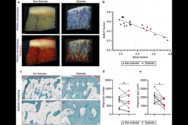
AbstractObjective Pathological bone formation requires energy derived from metabolic substrates including glucose, amino acids, and fatty acids. As bone marrow adipose tissue (BMAT) is a rich local source of fatty acids and is progressively depleted in osteoarthritic subchondral bone, we investigated whether lipolytic lipid mobilization is associated with osteogenic activity in sclerotic regions and whether this process is pharmacologically modifiable. Design Human knee osteoarthritis subchondral bone samples (n = 23) were analyzed using transcriptomic, histological, and ex vivo explant approaches. Associations between lipolytic and bone formation markers were assessed, with particular focus on PNPLA2, encoding adipose triglyceride lipase (ATGL), and alkaline phosphatase (ALP). Immunohistochemistry was used to localize lipolytic activation in relation to sclerotic bone regions. Ex vivo explants were used to assess adipocyte morphology, secretion of lipolysis products, including free fatty acids (FFA) and glycerol, and bone formation markers including ALP and P1NP. Pharmacological modulation of lipolysis was performed using forskolin to stimulate and metformin to inhibit lipid mobilization. Results Transcriptomic analysis revealed a strong association between ALPL expression and PNPLA2, suggesting a link between bone formation and lipolytic activity. Histological analysis confirmed local activation of ATGL in sclerotic subchondral bone regions. In explant cultures, reduced adipocyte size was accompanied by increased release of free fatty acids and glycerol, together with elevated ALP and P1NP levels. FFA secretion strongly correlated with ALP, supporting a relationship between BMAT lipid mobilization and osteogenic activity. PLIN1, a marker of lipid droplet integrity, was downregulated in isolated BMAT adipocytes but upregulated in paired stromal-vascular cells, suggesting transfer or utilization of BMAT-derived lipids by neighboring bone-forming cells. Pharmacological modulation confirmed that BMAT lipolysis is dynamically regulated: forskolin enhanced free fatty acid release, whereas metformin reduced it. However, short-term modulation did not significantly alter bone formation indices. Conclusion These findings identify BMAT lipolysis as a metabolic feature of subchondral bone sclerosis in knee OA. Mobilization of lipids from marrow adipocytes may provide energetic substrates that support osteoblast activity and pathological bone formation. BMAT lipolysis therefore represents a potentially modifiable metabolic pathway within the OA subchondral bone niche.