Multi-region calcium imaging in freely behaving mice with ultra-compact head-mounted fluorescence microscopes
Multi-region calcium imaging in freely behaving mice with ultra-compact head-mounted fluorescence microscopes
Xue, F.; Li, F.; Zhang, K.-m.; Ding, L.; Wang, Y.; Zhao, X.; Xu, F.; Zhang, D.; Sun, M.; Lau, P.-M.; Zhu, Q.; Zhou, P.; Bi, G.-Q.
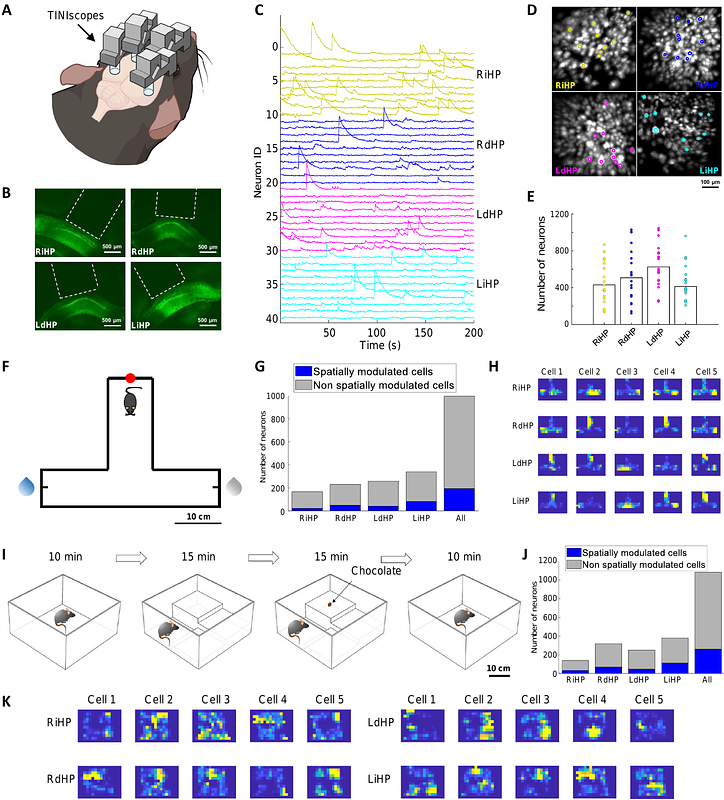
AbstractTo investigate the circuit-level neural mechanisms of behavior, simultaneous imaging of neuronal activity in multiple cortical and subcortical regions is highly desired. Miniature head-mounted microscopes offer the capability of calcium imaging in freely behaving animals. However, implanting multiple microscopes on a mouse brain remains challenging due to space constraints and the cumbersome weight of equipment. Here, we present TINIscope, a Tightly Integrated Neuronal Imaging microscope optimized for electronic and opto-mechanical design. With its compact and lightweight design of 0.43 g, TINIscope enables unprecedented simultaneous imaging of behavior-relevant activity in up to four brain regions in mice. Proof-of-concept experiments with TINIscope recorded over 1000 neurons in four hippocampal subregions and revealed concurrent activity patterns spanning across these regions. Moreover, we explored potential multi-modal experimental designs by integrating additional modules for optogenetics, electrical stimulation or local field potential recordings. Overall, TINIscope represents a timely and indispensable tool for studying the brain-wide interregional coordination that underlies unrestrained behaviors.