Unifying Orientation-Dependent Relaxation and Diffusion Around Axonal Fibers from DTI: Characterizing Fiber-Tract-Specific Anisotropic R2 Profiles of the Corpus Callosum
Unifying Orientation-Dependent Relaxation and Diffusion Around Axonal Fibers from DTI: Characterizing Fiber-Tract-Specific Anisotropic R2 Profiles of the Corpus Callosum
Pang, Y.; Raja, R.; Reddick, W. E.
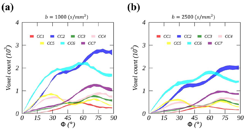
AbstractThis work aims to characterize fiber-tract-specific orientation-dependent R2 from the seven callosal segments (CC1-CC7) based on human brain Connectome high-resolution DTI datasets. WM voxels from each segment were masked by the thresholds of FA and mode of anisotropy. Encoded in T2W images (b-value = 0), an orientation-dependent R2 profile was constructed based on voxel-wise fiber orientations and characterized by a cone model. This allowed R2a, an orientation-dependent or anisotropic R2, to be separated from its orientation-independent or isotropic counterpart. Except for R2a, no discernible differences were found for the fits between the two b-values. On average, R2^a increased from 2.4{+/-}0.2 (1/s) to 3.2{+/-}0.3 (1/s) as the b-value increased. Furthermore, R2^a showed an increasing trend from CC1 to CC7 and the open angle alpha; of the cone fluctuated around 65{degrees}. As usual, FA was found increasing from CC1 to CC7 and from a higher to a lower b-value. In conclusion, we have shown that fiber-tract-specific anisotropic R2 profiles from the callosal segments can be characterized by a cone model. The proposed method offers a unique opportunity to reevaluate existing clinical DTI studies and optimize new ones for characterizing specific WM tracts in both healthy and diseased subjects.