Sialoglycoconjugate Profiling of Human Choroid, Retinal Pigment Epithelium, and Macular Degeneration Related Lesions
Sialoglycoconjugate Profiling of Human Choroid, Retinal Pigment Epithelium, and Macular Degeneration Related Lesions
Navratil, E. M.; Wenzel, P. A.; Flamme-Wiese, M. J.; Miller, J. E. B.; Wiley, L. A.; Stone, E. M.; Tucker, B. A.; Mullins, R. F.
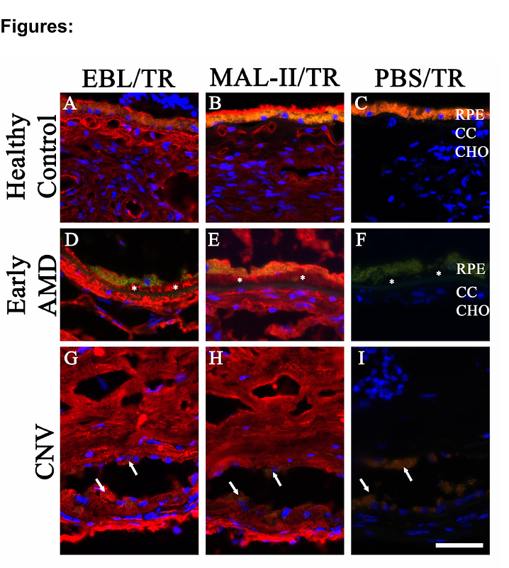
AbstractAge-related macular degeneration is a leading cause of central vision loss in the elderly. Early hallmarks of the disease include basal laminar deposit and choriocapillaris degeneration. The location and composition of sialoglycoconjugates in healthy and diseased choroid and disease-related lesions have not been thoroughly examined. This study utilized lectins to examine sialoglycoconjugates in human tissue, specifically Sambucus nigra/Elderberry Bark Lectin (EBL) and Maackia amurensis lectin II (MAL-II), to examine a-2,6 and a-2,3 sialic acids, respectively. EBL and MAL-II both label the choroid and basal laminar deposit, with slightly different patterns. Whereas MAL-II predominantly labels the choriocapillaris endothelium, EBL also labels Bruchs membrane and extracellular domains surrounding the vasculature (intercapillary pillars). EBL labeling overlaps with the distribution of complement factor H to a greater extent than MAL-II. After treatment with neuraminidase to remove terminal sialic acids, a battery of lectins was applied to sections of choroids. Lectins that recognize b-galactose, N-acetyllactosamine, galactose (b-1,3) N-acetylgalactosamine, and a- or b- N-acetylgalactosamine showed increased reactivity, including increased labeling of glycans in basal laminar deposits. This study provides insight into the location and partial identities of sialoglycoconjugates in the human choroid, with possible implications for the pathogenesis of macular degeneration.