Astrocyte-driven small vessel disease is an early, amyloid-independent feature of PSEN1 E280A familial Alzheimer's disease

Astrocyte-driven small vessel disease is an early, amyloid-independent feature of PSEN1 E280A familial Alzheimer's disease
Villalba-Moreno, J. L.; El-Amri, Y.; Kim, K.-Y.; Villalba-Moreno, N. D.; Shafiq, M.; Ortiz-Cordero, C.; Wang, S.; Ossa, J. A.; Suarez-Uribe, I.; Cardona-Madrigal, D.; Villegas, A.; Glatzel, M.; Krasemann, S.; Posada-Duque, R.; Kiessling, L. L.; Lopera, F.; Arboleda-Velasquez, J.; Kalaria, R. N.; Ellisman, M.; Sepulveda-Falla, D.
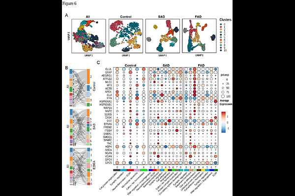
AbstractCerebral Small vessel disease (cSVD) is a prevalent feature of Alzheimer's disease (AD) pathology. Whether this pathology is a late consequence of amyloid and tau accumulation or an early, direct effect of PSEN1 dysfunction has remained unresolved. We found that it is more severe in familial AD (FAD) caused by E280A mutation in presenilin 1 (PSEN1). These cases present with a distinctive proteomic signature, associated with pathological features, more dysregulated in the occipital cortex (OC) compared to the frontal cortex (FC), and characterized by multiple dysregulated proteins involved in extracellular matrix (ECM) and RNA-associated processes. This proteomic fingerprint was associated with abnormal collagen build up, ECM disorganization, and signatures of aberrant angiogenesis. Six months old transgenic knock-in mice homozygous for Psen1 E280A mutation (PSEN1Ki) also showed a similar phenotype with microvascular tortuosity and proteomic changes. Critically, these mice develop neither A{beta} plaques nor tau tangles, indicating that the shared microvascular and RNA-associated changes are direct consequences of PSEN1 dysfunction rather than downstream effects of amyloid pathology. Remarkably, dysregulated RNA-associated protein networks overlapped between FAD and PSEN1Ki mice. Cerebral microvessels microstructure in PSEN1Ki mice at two months and six months showed abnormal astrocytic end-feet with lamellar deposits implicating blood-brain barrier damage. Finally, single nuclei transcriptomic analysis of AD patients and controls showed similar abnormal astrocytes in both sporadic and familial variants, but FAD astrocytes expressed dysregulated genes identified in the proteomic analyses, such as GLUL, APOE, and CLU. Our findings suggest that cSVD is an early pathological event in PSEN1 FAD and that is driven by abnormal RNA-associated processes and astrocytic dysfunction.