Accurate Automated Segmentation of Autophagic Bodies in Yeast Vacuoles Using Cellpose 2.0
Accurate Automated Segmentation of Autophagic Bodies in Yeast Vacuoles Using Cellpose 2.0
Marron, E. C.; Backues, J.; Ross, A. M.; Backues, S. K.
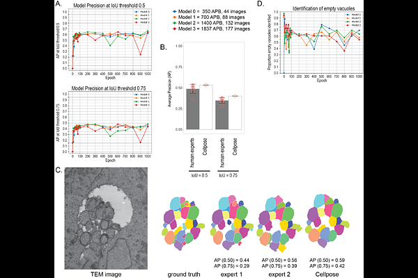
AbstractSegmenting autophagic bodies in yeast TEM images is a key technique for measuring changes in autophagosome size and number in order to better understand autophagy. Manual segmentation of these images can be very time consuming, particularly since hundreds of images are needed for accurate measurements. Here we describe a validated Cellpose 2.0 model that can segment these images with accuracy comparable to that of human experts. This model can be used for fully automated segmentation, eliminating the need for manual body outlining, or for model-assisted segmentation, which allows human oversight but is still five times as fast as the current manual method. The model and instructions for its use are presented here for the autophagy community.