Simulating magnetic rotational spectroscopy: a novel approach to intracellular rheology of adherent cells
Simulating magnetic rotational spectroscopy: a novel approach to intracellular rheology of adherent cells
Moore, C. P.; Ghasemi, F.; Berret, J.-F.
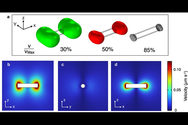
AbstractRecent studies have highlighted intracellular viscosity, alongside whole-cell elasticity, as a key biomechanical property with potential as a biomarker for cancer cell metastasis. Within the framework of cellular mechanobiology, magnetic rotational spectroscopy (MRS), which employs rotating magnetic wires to probe cytoplasmic rheology, has emerged as an effective method for quantifying intracellular viscoelasticity. This study examines microrheology data from three breast epithelial cell lines, MCF-10A, MCF-7, and MDA-MB-231, along with new data from HeLa cervical cancer cells. Here, MRS is combined with finite element simulations to characterize the flow field induced by wire rotation in the cytoplasm. COMSOL simulations performed at low Reynolds numbers show that the flow velocity is highly localized around the wire, and display characteristic dumbbell-shaped profiles. For wires most representative of MRS cell experiments, the shear rates are such that their product with the cytoplasmic relaxation time remains below 1, ensuring a linear flow regime. This outcome confirms that MRS can reliably measure the static viscosity of the intracellular medium in living cells. Nonlinear mechanical responses are predicted only in rare cases involving very short wires. This study demonstrates that integrating MRS intracellular measurements with COMSOL simulations significantly improves the reliability of in vitro assessments of cytoplasmic mechanical properties.