Whole-Brain, Region-Specific Astrocyte Reactivity and Morphological Remodeling After Diffuse Traumatic Brain Injury in A Gyrencephalic Ferret Model

Whole-Brain, Region-Specific Astrocyte Reactivity and Morphological Remodeling After Diffuse Traumatic Brain Injury in A Gyrencephalic Ferret Model
Bagherian, A.; Perez, C.; Kosub, A.; Chalijah Ysabelle Gonzales, R.; Patterson, A.; Bieniek, K. F.; Seidi, M.; Memar, M.
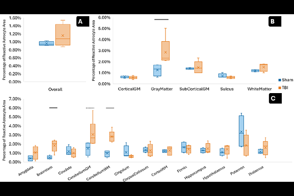
AbstractTraumatic brain injury (TBI) triggers pathological cascades that evolve across acute, subacute, and chronic phases. Astrocytes play a central role across these phases, and astrocyte reactivity is commonly evaluated using glial fibrillary acidic protein (GFAP) immunolabeling. However, in many TBI studies GFAP changes are characterized qualitatively or with manual or simple threshold-based measures on a small set of sections, limiting throughput and constraining analysis of region-specific heterogeneity in astrocyte responses. To overcome these limitations, we employed a ferret model of diffuse TBI (5 TBI, 5 sham), leveraging the ferret's gyrencephalic cortex, human-like regional fractional brain volumes, and astrocyte features that more closely resemble the human brain than rodent models. An AI-driven segmentation model validated for GFAP-stained ferret histology was integrated with atlas-based mapping to achieve whole-brain, region-resolved quantification of astrocyte reactivity over an average of 10 coronal slices per animal. Morphometric analysis using a custom SMorph-based pipeline characterized branching complexity and spatial domain features across defined regions. At seven days post-injury, TBI animals showed elevated astrocyte reactivity and hypertrophic remodeling, with significant expansion of convex hull area and elongation of secondary branches at the whole-brain level, most pronounced in the atlas-defined gray-matter region and cerebellum and brain-stem subregions, whereas white-matter showed a similar but less marked trend. Morphological changes were also detected in the hippocampus that did not show significant increases in astrocyte reactivity, indicating that structural remodeling represents a partially independent dimension of the astroglial response. These regional patterns are consistent with expected large tissue deformation and axonal strain in brainstem-cerebellar pathways and gray-matter at gray-white junctions in sagittal rotation, motivating future computational studies to quantify these links more directly. By combining region-resolved GFAP mapping with large-scale morphometry, this work provides a scalable framework for region-specific astrocyte mapping to support future multimodal, computational, and targeted neuroprotective studies.