Visible Light Optical Coherence Tomography Reveals Aging at the RetinalPigment Epithelium-Bruch's Membrane Interface
Visible Light Optical Coherence Tomography Reveals Aging at the RetinalPigment Epithelium-Bruch's Membrane Interface
Meng, R.; Kenney, R. C.; Pan, M.; Gupta, A. K.; Modi, Y. S.; Chauhan, P.; Curcio, C. A.; Srinivasan, V. J.
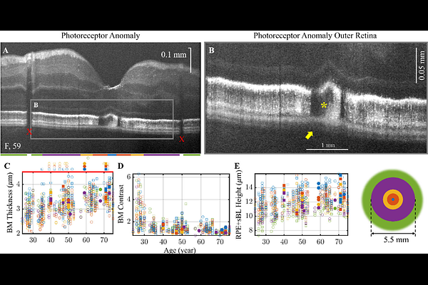
AbstractLandmark histological studies have shown that as the retina ages, lipids and other debris accumulate within Bruch's membrane (BM) and in spaces introduced between BM and the retinal pigment epithelium (RPE). These deposits grow with age, increasing the risk of age-related macular degeneration (AMD), the leading cause of irreversible vision loss for older adults globally. Current in vivo imaging lacks specificity to study BM and the important spaces at the RPE/BM interface in living human eyes, while histological techniques suffer from processing artifacts that distort photoreceptors. Here we employ visible light Optical Coherence Tomography (OCT), with 1 micrometer depth resolution, to quantitatively analyze these tissues in living eyes. We identify age-related changes in a human cohort without retinal pathology: thickening and loss of contrast of the hyper-reflective BM band, and thickening of the RPE together with the sub-RPE basal laminar space (RPE+sBL). Both forms of thickening were locally coupled depending on eccentricity, suggesting related biosynthetic mechanisms. A thicker BM and RPE+sBL were locally associated with anomalies in the overlying photoreceptors. Thus, sub-clinical changes in aging eyes detected by visible light OCT resemble early versions of deposits found in AMD. Visible light OCT depicts the relationship between RPE+sBL, BM, and photoreceptors in aging, holding promise to precisely and non-invasively grade ocular phenotypes ranging from normal aging to early AMD.