Mechanism of giant magnetic field effect in fluorescence of mScarlet3, a red fluorescent protein

Mechanism of giant magnetic field effect in fluorescence of mScarlet3, a red fluorescent protein
Xiang, K. M.; Lampson, H.; Hayward, R. F.; York, A. G.; Ingaramo, M.; Cohen, A. E.
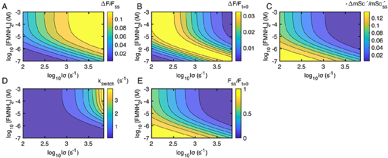
AbstractSeveral fluorescent proteins, when expressed in E. coli, are sensitive to weak magnetic fields1. We found that mScarlet3 fluorescence in E. coli reversibly decreased by 21% in the presence of a 60 mT magnetic field, the largest magnetic field effect (MFE) reported in any fluorescent protein. Purified mScarlet3 did not show an MFE, but addition of flavin mononucleotide (FMN) and simultaneous illumination with blue and yellow light restored the MFE. Through extensive photophysical experiments, we developed a quantitative model of the giant MFE in mScarlet3-FMN mixtures. The key reaction step involved electron transfer from fully reduced FMNH2 to triplet-state mScarlet3, to form a triplet spin-correlated radical pair. The magnetic field then controlled the branching ratio between singlet recombination vs. triplet separation. Our quantitative model of the mScarlet3-FMN photocycle provides a framework for design and optimization of magnetic field-sensitive proteins, opening possibilities in fluorescent protein-based magnetometry, magnetic imaging, and magnetogenetic control.