3D vascular quantitation with application to computational modeling: a pre-clinical light sheet microscopy, high resolution ultrasound, nano-computed tomography comparison study
3D vascular quantitation with application to computational modeling: a pre-clinical light sheet microscopy, high resolution ultrasound, nano-computed tomography comparison study
Zhang, D.; Lindsey, S. E.
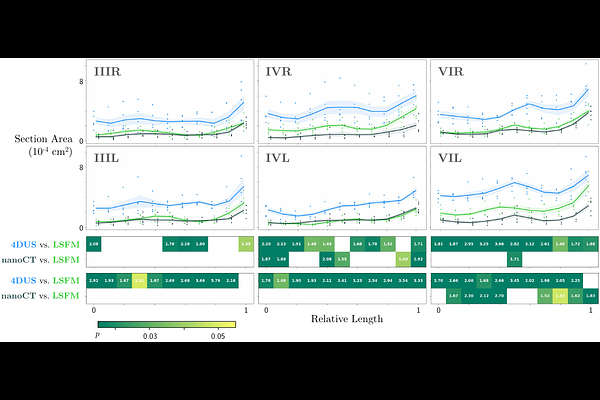
AbstractIt is increasingly necessary to both study biology in 3D and obtain quantitative measurements. Not all 3D-reconstructions are created equal, particularly when using the anatomical model as a basis for force calculations, i.e. computational modeling. Here, we compare 3D anatomical reconstructions from two emerging imaging modalities: 4D ultrasound (4DUS) and light sheet fluorescent microscopy (LSFM) against our previous nano-computed tomography (nanoCT) cohort data, using the tortuous highly intricate pharyngeal arch artery system of the chick embryo as a test bed. We highlight modality-specific morphological image acquisition discrepancies and their influence on subsequent computational fluid dynamics results. Overall, LSFM accurately captured quantitative volumetric measurements of small rapidly-changing vascular morphologies while 4DUS systematically inflated small tortuous vessels. Differences in image-based morphology changes led to significant changes in computationally-obtained force magnitudes and flow patterns linked to vessel angle and tortuosity. This validates LSFM as a comparative preclinical vascular quantitative imaging tool and suggests that 4DUS needs extensive 3D anatomical validation for non cardiac chamber vessels