Collagen fibre organisation revealed with computational scattered light imaging and polarimetric second harmonic generation microscopy
Collagen fibre organisation revealed with computational scattered light imaging and polarimetric second harmonic generation microscopy
Ettema, L.; Mazeika, V.; Alizadeh, M.; Abbasi, H.; Barzda, V.; Menzel, M.
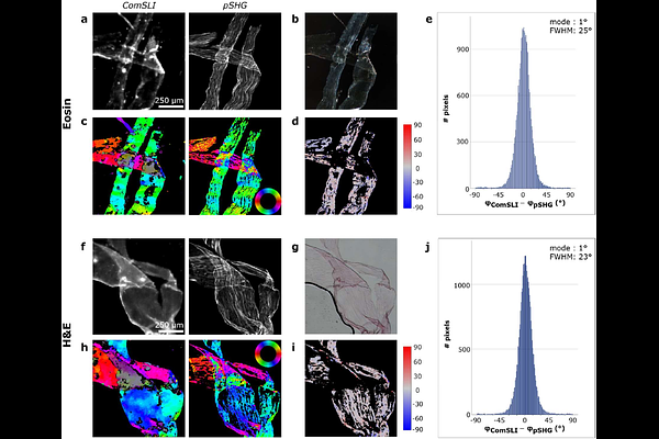
AbstractCollagen forms dense fibre networks in the human body, with the organisation directly influencing tissue mechanics and function in health and disease. A good understanding of this relation requires proper imaging techniques for visualising the dense collagen network. Previously, computational scattered light imaging was employed as a fast and easy-to-implement technique to retrieve the orientation of multi-directional collagen fibres, but results were not yet validated quantitatively. In this study, we validate the in-plane orientations of collagen fibres determined with computational scattered light imaging by performing comparative measurements with polarimetric second harmonic generation microscopy on rat tendon and bone sections. For rat tendon, sections with and without hematoxylin-and-eosin staining, folded tendon layers, and obliquely cut sections were investigated. Similar fibre orientations were obtained with both techniques in both tissues, with the highest degree of similarity found for in-plane, unidirectional fibres in the tendon sections. The techniques were able to retrieve the orientations of multi-directional crossing collagen fibres in folded rat tendon layers, and results were found to be unaffected by staining. While polarimetric second harmonic generation microscopy provides high resolution and ultrastructural information on collagen, computational scattered light imaging provides large field of view measurements with micrometre resolution.