Cytoplasmic Viscosity is a Potential Biomarker for Metastatic Breast Cancer Cells
Cytoplasmic Viscosity is a Potential Biomarker for Metastatic Breast Cancer Cells
Dessard, M.; Manneville, J.-B.; Berret, J.-F.
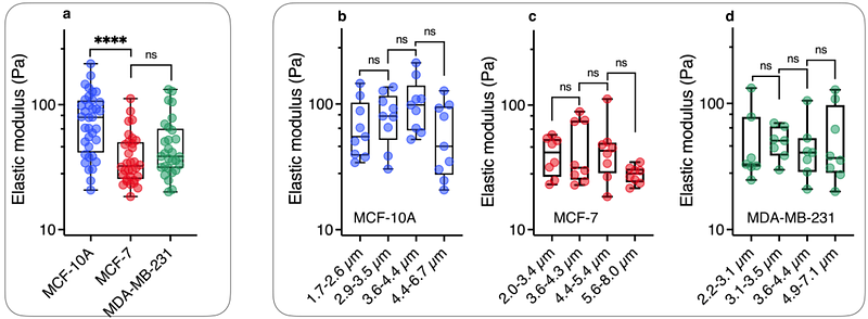
AbstractCellular microrheology has shown that cancer cells with high metastatic potential are softer compared to non-tumorigenic normal cells. These findings rely on measuring the apparent Young modulus of whole cells using primarily atomic force microscopy. This study aims to ex-plore whether alternative mechanical parameters have discriminating features with regard to metastatic potential. Magnetic rotational spectroscopy (MRS) is employed in the examination of mammary epithelial cell lines: MCF-7 and MDA-MB-231, representing low and high meta-static potential, alongside normal-like MCF-10A cells. MRS utilizes active micron-sized mag-netic wires in a rotating magnetic field to measure the viscosity and elastic modulus of the cy-toplasm. All three cell lines display viscoelastic behavior, with cytoplasmic viscosities ranging from 10-70 Pa s and elastic moduli from 30-80 Pa. It is found that the tumorigenic MCF-7 and MDA-MB-231 cells are softer than the MCF-10A cells, with a twofold decrease in elastic modu-lus. To differentiate cells with low and high malignancy however, viscosity emerges as the more discriminating parameter, as MCF-7 exhibits a 5 times higher viscosity as compared to MDA-MB-231. These findings highlight the sensitivity of cytoplasmic viscosity to metastatic potential, suggesting its potential utility as a mechanical marker for malignant cancer cells.