Automated Quantitative Assessment of Elastic Fibers in Verhoeff-Van Gieson-Stained Mouse Aorta Histological Images
Automated Quantitative Assessment of Elastic Fibers in Verhoeff-Van Gieson-Stained Mouse Aorta Histological Images
Lefebvre, A. E. Y. T.-S.; Mullis, M.
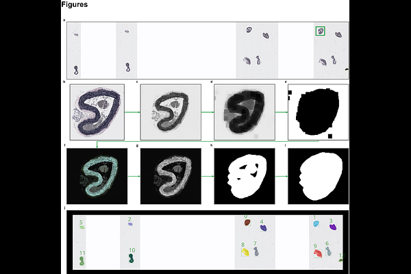
AbstractThe mechanical resilience of the aortic wall hinges on the organisation of its concentric elastic laminae, yet histological evaluation of these fibers remains largely qualitative and observer-dependent. We present a fully automated, stain-aware pipeline that transforms Verhoeff-Van Gieson (VVG) whole-slide images of mouse aortae into reproducible, quantitative maps of elastic-fiber architecture. Leveraging optical-density deconvolution to disentangle elastin from collagen, the workflow couples multi-resolution processing with graph-based skeletonisation to preserve gigapixel detail while scaling efficiently. It returns pixel-level measurements of fiber thickness, tortuosity, lamina count and network complexity, together with validation snapshots for transparent quality control. By eliminating observer bias and delivering high-throughput morphometry, our framework enables powered genotype-phenotype screens in genetically diverse mouse populations and provides objective read-outs for interventions aimed at preserving matrix integrity. The modular codebase is open-source, readily extendable to other elastin-rich tissues or stains, and forms a bridge between qualitative microscopy and biomechanical phenotyping-setting the stage for large-scale, data-driven exploration of vascular structure-function relationships.