Spatially resolving how phosphorylation affects β-cardiac myosin activity in porcine myofibril sarcomeres with single molecule resolution


Spatially resolving how phosphorylation affects β-cardiac myosin activity in porcine myofibril sarcomeres with single molecule resolution
Pilagov, M.; Steczina, S.; Naim, A.; Regnier, M.; Geeves, M.; Kad, N. M.
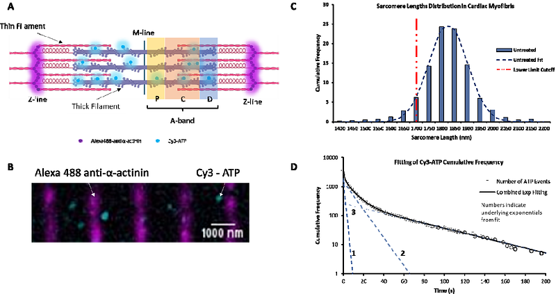
AbstractCardiac muscle contraction is mediated by myosin binding from the thick filament of the sarcomere to the thin filament in an ATP powered reaction. This process is highly regulated on a beat-to-beat basis by calcium interactions with the thin filament. Additionally, the number of heads available for participation in contraction is also regulated, resulting in a dynamically variable reserve of heads for controlling contractile force. We aimed to discover the size of this reserve and how it is modulated by phosphorylation. Using single molecule imaging of fluorescently labelled ATP molecules binding and releasing myosins within porcine cardiac sarcomeres, we could determine myosin activity with high spatial resolution. We find three kinetic species when examining the myosin ATPase. The fastest is consistent with non-specific ATP binding to myosins surface, and the slower two species are consistent with the previously identified DRX and SRX states. The former is thought to represent myosins in an ON state, ready to interact with the thin filament and the latter an OFF state with slowed ATPase that constitutes the cardiac reserve. We find that the cardiac reserve is 50% in the sarcomere and this can be sub-divided into the P-, C- and D-zones, with the D-zone having the least population of OFF heads (44%). Treatment with PKA phosphorylates cardiac myosin binding protein-C (cMyBP-C) leading to a 16% reduction in reserve in the C-zone (where cMyBP-C is found), a 10% reduction in the P-zone, and an unexpected 8% increase in the D-zone. By contrast, myosin regulatory light chain (RLC) phosphorylation with myosin light chain kinase (MLCK) resulted in a large 24% decrease in reserve myosins, interestingly the least affected area of the sarcomere was the C-zone. Altogether these data suggest that cMyBP-Cs interaction with RLC governs the degree of activation due to RLC phosphorylation.