Spatial Clustering Analysis with Spectral Imaging-based Single-Step Multiplex Immunofluorescence (SISS-mIF)


Spatial Clustering Analysis with Spectral Imaging-based Single-Step Multiplex Immunofluorescence (SISS-mIF)
Nakamura, T.; Kaneko, N.; Taguchi, T.; Ikeda, K.; Sakata, M.; Inoue, M.; Kuwayama, T.; Tatsuta, H.; Onishi, I.; Kurata, M.; Nakagawa, K.
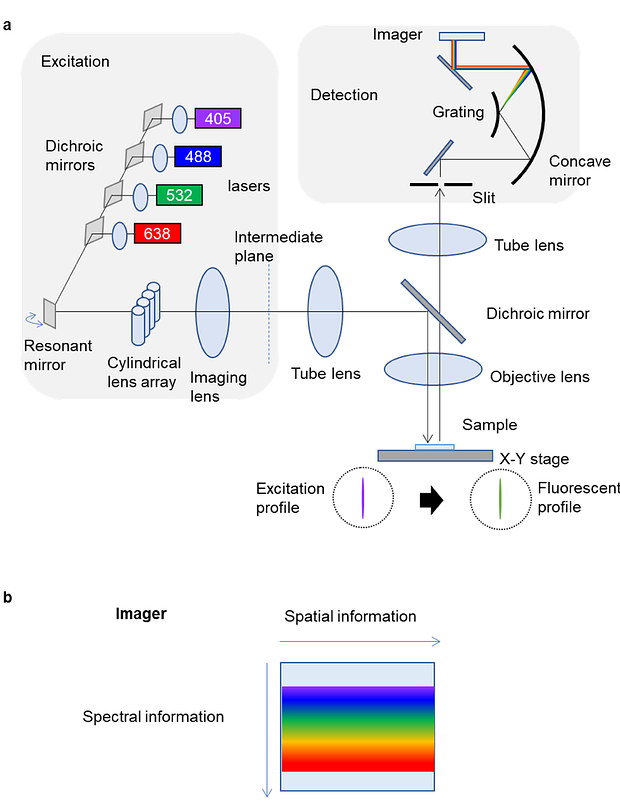
AbstractPrecision medicine, anchored in spatial biology, is essential for the accurate diagnosis of cancer and prediction of drug responses. We have introduced the Spectral Imaging-based Single-Step Multiplex Immunofluorescence (SISS-mIF) technique, which leverages hyperspectral imaging to simultaneously capture fluorescence spectra. This approach automatically optimizes tissue autofluorescence spectra for each image, facilitating the use of fluorescent direct-labeled antibodies for multicolor staining in a single step. Unlike conventional methods, images are outputted as antibody counts rather than fluorescence intensity, allowing for consistent comparisons under different imaging conditions. We demonstrate that this technique allows for identical cell detection of CD3, CD5, and CD7 in T-cell lymphoma on a single slide. The utilization of fluorescent direct-labeled antibodies enables the triple staining of CD3, CD5, and CD7 without cross-reactivity, maintaining the same intensity as single stains. Moreover, we developed a joint Non-Negative Matrix Factorization-based Spatial Clustering Analysis (jNMF-SCA) with a modified spectral unmixing system, highlighting its potential as a supportive diagnostic tool for T-cell lymphoma.