QUANTIFYING GLYCOGEN AND LIPID DROPLET SYNTHESIS IN OVARIAN AND CERVICAL CANCER CELLS USING DEUTERATED RAMAN PROBES WITH STIMULATED RAMAN SCATTERING MICROSCOPY
QUANTIFYING GLYCOGEN AND LIPID DROPLET SYNTHESIS IN OVARIAN AND CERVICAL CANCER CELLS USING DEUTERATED RAMAN PROBES WITH STIMULATED RAMAN SCATTERING MICROSCOPY
Pierson, R. N.; Gupta, S. A.; Zhang, M.; Kaiser, L. C.; Tumey, L. N.; Lu, F.
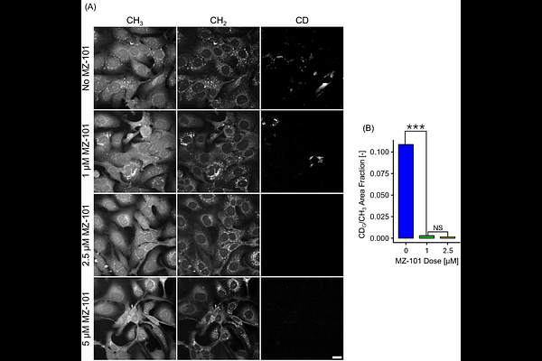
AbstractEpithelial ovarian cancer remains one of the most lethal malignancies among women, with late-stage diagnoses yielding 5-year survival rates below 30%. The metabolic heterogeneity of the tumor microenvironment (TME) highlights the need for methods capable of rapid, chemically specific phenotyping. Stimulated Raman scattering (SRS) microscopy when combined with deuterium labeled metabolites enables the non-invasive high contrast interrogation of cellular metabolic pathways. In this study, we used SRS microscopy to profile fatty acid and glycogen metabolism in epithelial ovarian cancer (SKOV-3) and cervical cancer (HeLa) cell models. Deuterium labeled glucose revealed striking differences in glycogen synthesis and intracellular distribution, with SKOV-3 cells exhibiting markedly greater single-cell heterogeneity than HeLa. Complementary measurements of lipid droplet (LD) synthesis and turnover under nutrient starvation further revealed cell-line-specific metabolic strategies, identifying LD and glycogen dynamics as a potential diagnostic marker of cancer metabolic phenotypes. These results demonstrate that SRS microscopy in the Raman silent region, paired with metabolic labeling, can sensitively resolve metabolic diversity across cancer cell subpopulations. Such metabolic phenotyping may inform both early diagnostic strategies and therapeutic approaches that combine cytotoxic treatment with targeted metabolic disruption.