Rapid immunostaining and high-resolution three-dimensional light-sheet microscopy of intact calcified tissues

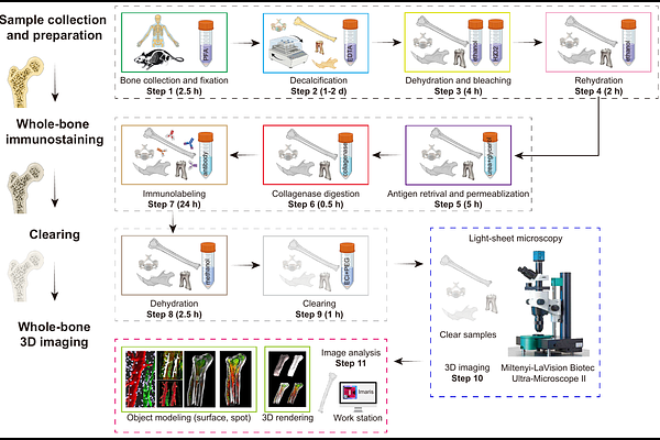
Rapid immunostaining and high-resolution three-dimensional light-sheet microscopy of intact calcified tissues
Ding, Z.; Shi, Y.; Liu, H.; Li, C.; Chen, J.; Cohen-Solal, M.; Kusumbe, A. P.
AbstractHigh-resolution 3D imaging is an important strategy for visualizing and analysing complex skeletal tissue architecture and the bone marrow microenvironment. However, multicolor immunolabeling and imaging of intact skeletal tissues are technologically challenging. The current immunolabeling and clearing methods for intact skeletal elements are very limited, time-consuming and generate low-resolution data or depend on the use of reporter mice. Here, we describe a protocol for efficient clearing and immunolabeling of intact calcified tissues that enables superfast, single-cell resolution, and quantitative 3D light-sheet imaging of intact skeletal elements and teeth. A key aspect of our protocol is the addition of a collagenase digestion step after fixation and decalcification. This step enhances antibody penetration, resulting in deep, comprehensive staining throughout immunostained bones and other calcified tissues. The protocol includes soft tissue removal, fixation, decalcification, bone dehydration, and bleaching, followed by antigen retrieval and permeabilization before the collagenase digestion step. This procedure is performed to prepare the samples for the tissue clearing process that improves bone tissue transparency prior to light-sheet imaging. The entire protocol, from bone collection to image analysis and quantification, takes about 4 days to complete, thus offering significant improvements over previous methods. This protocol is broadly applicable to the visualization of bone microstructure, bone marrow analysis, vascular and neural network mapping, and the study of signaling molecules in bone development and growth. The protocol requires experience with standard tissue processing and immunostaining techniques, and prior experience in tissue clearing and light-sheet imaging is beneficial but not essential. Key pointsO_LIA protocol for efficient clearing and immunolabeling of intact calcified tissues that enables superfast, high-resolution, and quantitative 3D imaging of various intact bones and teeth. C_LIO_LIThe entire protocol takes only 4 days to complete the comprehensive staining and perfect transparency throughout the intact bones, offering significant improvements over previous methods. C_LI Key referencesBiswas, L. et al. Cell 186, 382-397.e24 (2023): https://doi.org/10.1016/j.cell.2022.12.031