The 3D Ultrastructure of C. elegans Gut Granules
The 3D Ultrastructure of C. elegans Gut Granules
Archer, G.; Bartlowe, S.; Bayarbadrakh, B.; Bok, D.; Bushong, C.; Carroll, A.; Cole, M.; Dahl, E. J.; Denier, N.; Elewa, A.; El Harchi, H.; Fisher, D.; Grant, H.; Grinfeld, R.; Grijalva, A.; Hale, A.; Hendricks, C.; Iskandar, M.; Kagan, S.; Mistry, N.; Keuneke, H.; Lammers, L.; Lyons, A.; Maclin, Q.; Moore, T.; Munro, C.; Nickerson, G.; Papalia, B.; Peacock, K.; Ritzman, L.; Ross, A.; Samineni, R.; Scales, M.; Schotz, L.; Sikkema, W.; Slabaugh, T.; Sorenson, A.; Swisher, R.; Sutherland, S.; Valdes, D.; Ward, F.; Wojdyla, P.
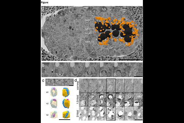
AbstractWe identify an endoderm-restricted organelle in published volume electron microscopy datasets of C. elegans embryos. The organelle consists of a tubular ring surrounding a membrane-bound compartment harboring a prominent dense particle and exhibits a basal polarity and size concordant with canonical gut granules. This finding offers ultrastructural detail to recent evidence that gut granules are bi-lobed organelles with two distinct compartments.