Mechanical Coordination of Intestinal Cell Extrusion by Supracellular 3D Force Patterns
Mechanical Coordination of Intestinal Cell Extrusion by Supracellular 3D Force Patterns
Matejcic, M.; Wang, M.; Lopez Serrano, E.; Perez-Gonzalez, C.; Houtekamer, R.; Ceada, G.; Roca-Cusachs, P.; Gloerich, M.; Trepat, X.
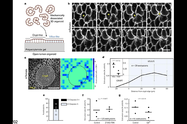
AbstractEvery day, the mammalian intestinal epithelium extrudes millions of cells to sustain tissue self-renewal. Despite its fundamental role in intestinal homeostasis, the mechanisms that trigger, compartmentalize, and execute intestinal cell extrusion remain largely unknown. Here, using intestinal organoids, we map the three-dimensional forces and cytoskeletal dynamics that drive intestinal cell extrusion. We show that, unlike in other epithelia, extrusion is initiated by the sudden dissolution of a contractile myosin 2A meshwork triggered by a calcium influx. Following meshwork dissolution, the extruding cell and its neighbors generate an upwards traction force that requires myosin contractility but is generated by lamellipodial protrusions in neighboring cells. Importantly, these lamellipodia not only act as force generators but also determine whether extrusion occurs apically or basally, serving as symmetry breakers of the process. Finally, we show that compartmentalization of cell extrusion to the outside of the intestinal crypt does not require curvature and instead depends on myosin 2A. Our findings reveal that the intestinal epithelium exhibits a distinctive mode of extrusion, in which tension differentials - rather than compressive stresses from crowding - trigger and compartmentalize cell removal.