3D imaging of the pregnant uterus reveals an extensively invasive mouse placenta requiring CXCL12-CXCR4 signaling
3D imaging of the pregnant uterus reveals an extensively invasive mouse placenta requiring CXCL12-CXCR4 signaling
Zwierzynski, J. B.; Moufarrej, M. N.; Red-Horse, K.
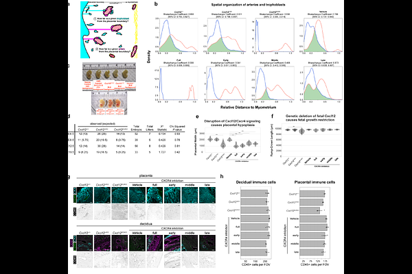
AbstractSuccessful pregnancy requires exquisite balance: the placenta must invade just enough to access maternal blood but not so deep it remains attached at birth. Disrupting this balance causes life-threatening pregnancy complications, for which treatments remain limited. Animal models are desperately needed to discover mechanisms underlying balanced uteroplacental development and how pregnancy complications arise, but this is hampered by the view that mouse placentation lacks human characteristics such as extensive trophoblast invasion and targeting of uterine spiral arteries. Here, we utilize 3D imaging, mouse genetics, and pharmacological perturbations to demonstrate that: (1) The mouse placenta invades more extensively than previously recognized with most spiral arteries heavily enveloped by fetal trophoblasts, (2) This process is disrupted without CXCL12-CXCR4 signaling specifically during early pregnancy, and (3) Disrupting early uteroplacental development ultimately results in excessively deep trophoblast invasion, closely mimicking the pregnancy complication placenta accreta. Mechanistically, uterine epithelium, stroma, and arteries activate CXCR4 signaling in early pregnancy, and inhibition causes decidualization failure, followed by dissolution of spiral artery development. Trophoblasts consequently migrate deep into uterine muscle and its arteries, reproducing hallmarks of human accreta. Thus, with 3D imaging, the mouse more effectively models human uteroplacental development and defines an early etiological window for intervention.