iPSC-derived skeletal muscle spheroids for Duchenne Muscular Dystrophy modeling
iPSC-derived skeletal muscle spheroids for Duchenne Muscular Dystrophy modeling
Esposito, J.; Leite, F. d. S.; Barbosa, I. N.; Martins, T. M. d. M.; Olberg, G. G. d. O.; Tanoury, Z. A.; Telles-Silva, K. A.; Pardo, M. C. d. S.; Jazedje, T.; Bortolin, R. H.; Hirata, M. H.; Pourquie, O.; Zatz, M.
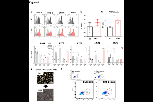
AbstractBackground The progressive skeletal muscle degeneration observed in Duchenne Muscular Dystrophy (DMD) patients requires multiple cycles of satellite cells (SCs) activation to promote tissue regeneration. Dystrophic SCs present intrinsic defects, and the disrupting fibrotic niche hinders appropriate muscle recovery. Traditional 2D culture systems face challenges in modeling the DMD muscle niche and SCs behavior. Our aim was to validate a 3D culture of skeletal muscle spheroids (iSMS) for DMD modeling, as compared to the traditional 2D culture, while investigating the pathophysiological mechanisms of dystrophin deficiency in vitro. Methods To compare iSMS with traditional 2D myogenic differentiation, we differentiated PAX7 reporter wild-type (WT), dystrophic (DMD) isogenic induced pluripotent stem cells (iPSCs), and patients iPSCs, characterized myogenic markers levels and assessed differences in proliferation and differentiation using RT-qPCR, immunofluorescence, and flow cytometry. Results Our data showed that although both 2D and iSMS culture systems generated myogenic progenitors positive for MYOD, MYOG, MYF5, and MYH3, iSMS improved PAX7 expression in vitro. Moreover, we identified a differential regulation of canonical Notch signaling genes between iSMS and 2D, which may influence the comparison between WT and DMD. We also characterized the differentiation of myogenic progenitors derived from 2D and iSMS towards elongated myofibers, providing a valuable comparison with muscle fibers differentiated from human primary myoblasts. Additionally, DMD iSMS derived progenitors proliferated at reduced levels compared with WT iSMS, a characteristic not observed in progenitors derived from 2D cultures. Finally, we performed iSMS and 2D myogenic differentiation of iPSC lines from three patients with DMD, thus validating the iSMS protocol for DMD modeling. Conclusion Our results highlight the important advantages of using the iSMS differentiation platform over 2D for disease modeling. Exploring these 3D systems may help to gain a deeper understanding of SCs behavior to advance in novel treatments for DMD, which might be applicable to other forms of muscular disorders.