A modified RGB trichrome (HemRGB) for improving nuclear staining: application to colorectal cancer histopathology

A modified RGB trichrome (HemRGB) for improving nuclear staining: application to colorectal cancer histopathology
Gaytan, F.
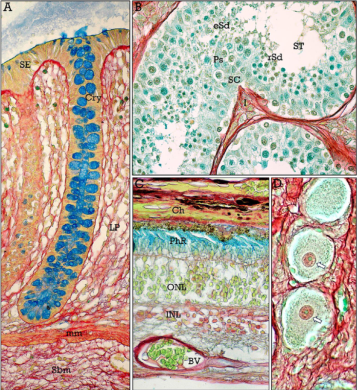
AbstractThe RGB trichrome staining method has been used to highlight two major components of the extracellular matrix, collagen and glycosaminoglycans. While the RGB trichrome efficiently stains extracellular matrix components, it lacks a nuclear stain, limiting its application in histopathology. To address this issue, a modification of the original stain, named HemRGB trichrome, has been developed. This modification incorporates iron hematoxylin for improving nuclear staining while retaining specificity for the staining of extracellular matrix. The application of HemRGB trichrome staining to samples from both normal colonic tissues and colorectal adenocarcinomas (CRC) provides a robust nuclear staining, together with a high-contrasted staining of tumor microenvironmental components, such as infiltrating immune cells, collagen and ground substance, extracellular mucins, as well as contrasted interfaces between CRC metastases and liver parenchyma. This study underscores the potential of HemRGB trichrome as a valuable tool for histopathological studies, especially for cancer evaluation, where nuclear characteristics are particularly relevant.