Ultrastructure analysis of mitochondria, lipid droplet and sarcoplasmic reticulum apposition in human heart failure
Ultrastructure analysis of mitochondria, lipid droplet and sarcoplasmic reticulum apposition in human heart failure
Latchman, N. R.; Stevens, T. L.; Bedi, K.; Prosser, B. L.; Margulies, K. B.; Elrod, J. W.
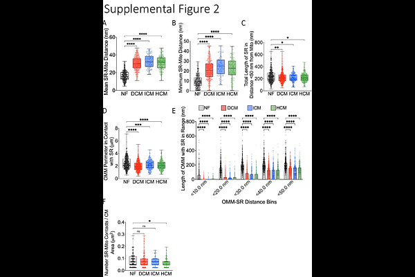
AbstractBackground: Cardiomyocyte structural remodeling is reported as a causal contributor to heart failure (HF) development and progression. Growing evidence highlights the role of organelle apposition in cardiomyocyte function and homeostasis. Disruptions in organelle crosstalk, such as that between the sarcoplasmic reticulum (SR) and mitochondria, are thought to impact numerous cellular processes such as calcium handling and cellular bioenergetics; two processes that are disrupted and implicated in cardiac pathophysiology. While the physical distance between organelles is thought to be essential for homeostatic cardiomyocyte function, whether the interactions and coupling of organelles are altered in human heart failure remains unclear. Methods: Here, we utilized transmission electron microscopy and careful quantification of ultrastructure to characterize the changes in organelle apposition in cardiomyocytes isolated from the hearts of patients diagnosed with various types of HF. Subsequently we employed molecular approaches to examine the expression of proposed organelle tethers. Results: We demonstrate that cardiomyocytes isolated from dilated cardiomyopathy, hypertrophic cardiomyopathy and ischemic cardiomyopathy hearts display smaller, more rounded mitochondria, as compared to nonfailing controls. Failing cardiomyocytes also exhibited disrupted SR-mitochondria juxtaposition and changes in the expression of proposed molecular tethers. Further analysis revealed alterations in lipid droplet dynamics including decreased lipid droplet content and less lipid droplets in association with mitochondria in failing cardiomyocytes. Conclusion: Here we observed changes in organelle dynamics in cardiomyocytes isolated from heart failure patients diagnosed with differing etiologies. Our results suggest that organelle structure and apposition may be a ubiquitous contributor to human HF progression.