Reconstructing the developmental history of the cortex from postmortem tissue

Reconstructing the developmental history of the cortex from postmortem tissue
Murakami, T. C.; Heintz, N.
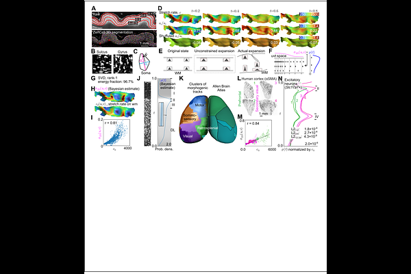
AbstractSingle-cell-resolution mapping of the human brain is central to understanding the link between cellular-level phenotypes and disease. Tissue clearing and mesoscale imaging have facilitated organ-wide quantification of cell populations and are expected to drive the identification of pathological deficits in neurological disorders. However, mesoscopic comparison of human brain tissue remains challenging because of large variability in cortical folding patterns and heterogeneous cell distribution. Here, we present a 'retrodictive' analysis of the cortex that reconstructs the proliferation history of neural stem cells from a static snapshot of tissue, providing a developmentally grounded coordinate system for comparison across samples. The analysis showed that most of the heterogeneity in cell distribution can be explained by the physical displacement caused by folding, which allows folding-induced variation to be removed and additionally reveals a possible mechanism of cortical folding. Together with a Bayesian framework, we estimated, with quantified uncertainty, the proliferation rate of a representative stem cell that summarizes the population of each cortical region. This analysis enables researchers to distill inter-individual differences down to stem cell properties and their distributions on the protomap. We anticipate our workflow to be a foundational framework for 3D neuropathological analysis, providing a common coordinate for the cortex-wide analysis of cells across individuals in development, aging, and disease.