Mycobacterium tuberculosis ESX-T7SS impacts the 3D architecture of tuberculous lesion in susceptible mice
Mycobacterium tuberculosis ESX-T7SS impacts the 3D architecture of tuberculous lesion in susceptible mice
Beltran, C. G. G.; Kriel, J.; Nolan, M. B.; Botha, S. M.; Ciccarelli, A.; Loos, B.; Gutierrez, M.; Walzl, G.
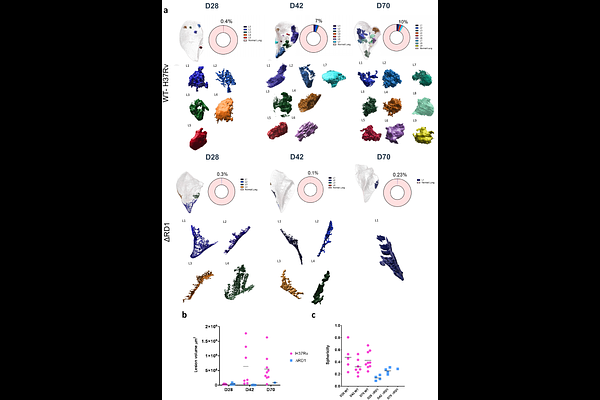
AbstractTuberculosis (TB) is characterized by the formation of heterogenous, immune-rich granulomas present in various forms in the lungs. Both host and pathogen contribute to this heterogeneity however the molecular and cellular drivers of within-host granuloma heterogeneity remain to be fully elucidated. This knowledge gap is due to a lack of experimental approaches that can fully capture the complex dynamics of the lung architecture, dynamics of host-pathogen interplay and pathogenesis. Here, we developed an approach that combines PACT-based clearing with light sheet fluorescent microscopy for visualizing lesion architecture development and lung involvement in M. tuberculosis-infected C3HeB/FeJ susceptible mice. This 3D modelling of whole lung lobes approach revealed critical architectural features in lesion development and lung involvement that were not apparent using traditional thin section imaging. Wild type M. tuberculosis infection triggered a clear and well-organized granulomatous-like organization with substantial growth throughout the infection period that invaded a high percentage of the total lung volume. In contrast, infection with the avirulent ESX-1 deletion mutant strain Mtb {Delta}RD1 showed an altered growth pattern with diffuse and sparsely organized CD11b recruitment to sites of infection. Moreover, most of the Mtb {Delta}RD1-triggered lesions were present in the periphery of the lungs and did not display any formal organization. We further provide a novel way of interrogating PACT-cleared tissue for high ultrastructural content using volumetric correlative light and electron microscopy, allowing individual immune cell populations to be tracked and their fate within the granuloma captured. Ultimately, the combination of both modalities allowed an unprecedented view of the architectural distribution of M. tuberculosis in the lungs and the progression of lesion development over time. Our data highlight that ESX-1 from M. tuberculosis is required for lesion architecture progression in a susceptible mouse model of TB.