Spatial analysis of Hofbauer cell transcriptome, distribution and morphology in placentas exposed to Plasmodium falciparum
Spatial analysis of Hofbauer cell transcriptome, distribution and morphology in placentas exposed to Plasmodium falciparum
Ataide, R.; Harding, R.; Dharmaratne, M.; Chen, Y.; Fielding, K.; Whitehead, L.; Rogers, K.; Anttila, C.; Ling, L.; Hickey, P.; Amann-Zalcenstein, D.; Moya, E.; Mhango, G.; Kamiza, S.; Randall, L.; Bennett, C.; Mzembe, G.; Mwangi, M. N.; Braat, S.; Phiri, K.; Pasricha, S.-R.
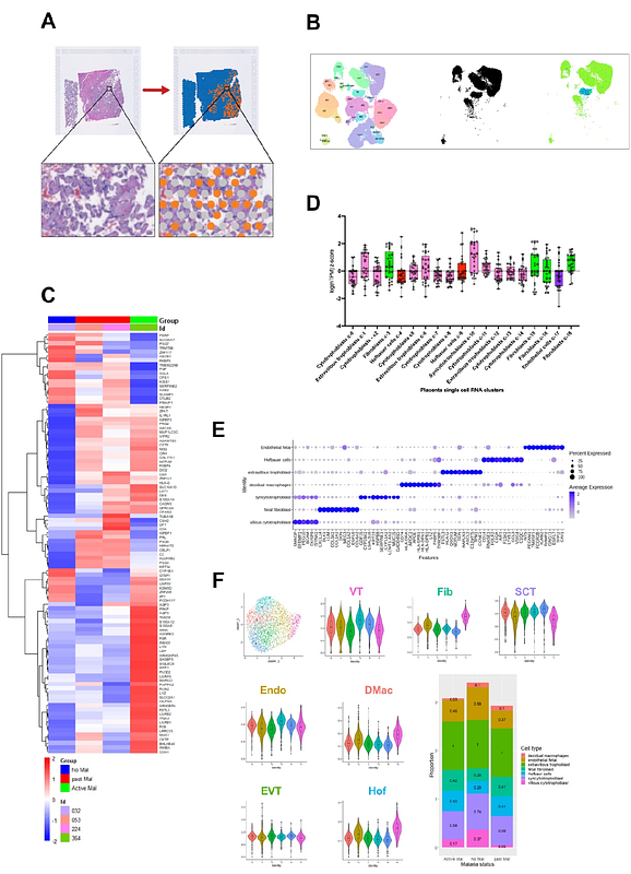
AbstractPlacental infection remains a significant health burden for mothers and their babies in low-income countries, especially in sub-Saharan Africa, where malaria transmission is intense. An increase in inflammatory biomarkers and poor vascularisation are characteristics of placentas infected with malaria. Hofbauer cells (HBCs) - placental villous macrophages of fetal origin - are one of the most abundant immune cells in the placenta. HBCs are thought to have roles in angiogenic processes and have been linked with the pathophysiology of several infections and inflammatory conditions during pregnancy, including malaria (caused by Plasmodium falciparum). However, there is limited in situ data on the transcriptional, proteomic or morphologic profile of these cells either during or following clearance of P. falciparum infection. We leveraged placental samples prospectively collected at delivery from 610 Malawian women enduring a high burden of malaria and other infections and nutritional deficiencies. We profiled placentas through spatial transcriptomic and proteomic platforms to discern in situ HBC features that could distinguish placentas with or without evidence of past malaria. In this cohort, past placental infection was common and was associated with lower birth weight babies (adjusted effect [95% confidence interval], -80.9 [-165.9, -3.7] g, P= 0.040). However, at term, HBC numbers, abundance, and transcriptional profiles from placentas with evidence of past infection were similar to those of placentas without malaria. HBCs may recover post-infection back to a basal state or may be replaced in the tissue over the course of pregnancy. Placentas with evidence of past malaria did show evidence of reduced fetal vessel development (mean area difference: -22.8% [-37.6, -7.9], P=0.003). Reduced vascular development following infection early in pregnancy may reflect disturbances to the normal vasculogenic and angiogenic processes, of which HBCs are an integral part.