Quantifying Intracellular Mechanosensitive Response upon Spatially Defined Mechano-Chemical Triggering
Quantifying Intracellular Mechanosensitive Response upon Spatially Defined Mechano-Chemical Triggering
Zare-Eelanjegh, E.; Lewis, R. T.; Lüchtefeld, I.; Kutay, U.; Zambelli, T.
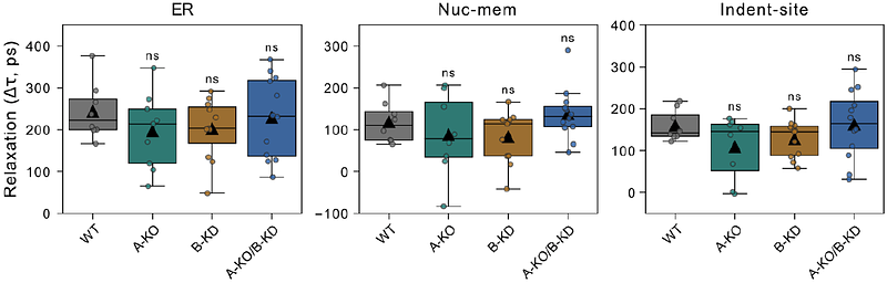
AbstractThe mechanotransduction process relies on the interaction of mechanical and biochemical cues, transmitting cellular forces to intracellular organelles to activate biochemical pathways and elicit responses. This involves mechanoresponsive components like actin filaments, microtubules (MTs), and the lamin meshwork. Fluidic force microscopy (FluidFM), a force-controlled micropipette allows for the manipulation of intact cells mechanically and chemically, providing a novel approach to study mechanotransmission in cells in situ. FluidFM combined with fluorescence lifetime imaging microscopy (FLIM), enables high-resolution mapping of intracellular tension dynamics. Here, we used cells with varying nuclear lamina compositions to explore the laminas role in initiating mechanoresponse to external cues. We found that A-type and B-type lamins trigger nuclear mechanoresponse distinctly, with A-type lamins contributing to nuclear elasticity, whereas B-type lamins influence viscous response. Moreover, MTs underwent mechanical adaptation and assisted in releasing the tension in lamin A/C knockout (KO) cells, contrasting with healthy cells where MTs aid to preserve the tension locally rather than transferring it. This research provides insights into the dynamic mechanoresponse of cellular components and supports targeted therapies for mechanical stress-related diseases.