Immunoglobulins are rapidly internalized by neurons after CNS injury and cleared through lysosomal degradation

Immunoglobulins are rapidly internalized by neurons after CNS injury and cleared through lysosomal degradation
Castellanos-Molina, A.; Boisvert, A.; Ferry, J.; Belanger, D.; Laroche, A.; Menet, R.; Crepeau, F.; Lessard, M.; Fortin, N.; Vallieres, N.; Allaeys, I.; Bertrand, N.; ElAli, A.; Boilard, E.; Lacroix, S.
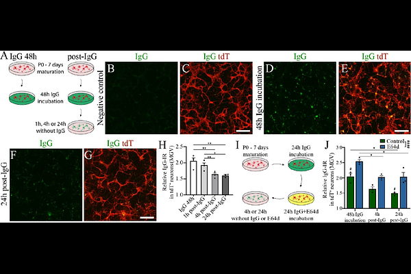
AbstractSpinal cord injury (SCI) causes hemorrhage and blood-spinal cord barrier disruption, allowing blood-derived molecules to infiltrate the parenchyma. While immunoglobulins (Ig) are abundant plasma proteins, their distribution and cellular targets within the injured spinal cord remain poorly defined. Here, we show that circulating non-autoimmune immunoglobulins rapidly infiltrate the spinal cord after injury in mice and disseminate beyond the lesion core. IgG, IgM, and IgA accumulate within the parenchyma early post-injury, with IgG displaying the widest spatial distribution, reaching distant spinal segments within hours. Neurons are the predominant cell type internalizing immunoglobulins in the gray matter, whereas astrocytes exhibit moderate uptake in white matter. Intra-cisterna magna administration of fluorescent serum-derived IgG reveals that neurons and astrocytes internalize IgG under physiological conditions, independently of Fc receptor engagement. Although the neonatal Fc receptor (FcRn) has minimal impact on CNS IgG recycling, its genetic deletion significantly improves locomotor recovery after SCI. Both in vitro and in vivo, neurons clear IgG through lysosomal degradation. Following SCI, inhibition of lysosomal proteases with the clinically approved drug E64d increases CNS IgG retention without compromising locomotor recovery. These findings establish neurons as key targets of circulating immunoglobulins after CNS injury and reveal IgG uptake and clearance pathways that may be leveraged to improve therapeutic performance of monoclonal antibody treatments.