Biogenesis and antigen presentation properties of dendritic cell-derived apoptotic bodies

Biogenesis and antigen presentation properties of dendritic cell-derived apoptotic bodies
Hodge, A. L.; Santavanond, J. P.; Shi, B.; Caruso, S.; Oveissi, S.; Vella, C.; Audi, O.; Ozkocak, D. C.; Rutter, S. F.; Phan, T. K.; Jiang, L.; Arakawa, S.; Shimizu, S.; Yoshino, I.; Atkin-Smith, G. K.; Ryan, G. F.; Chen, W.; Deng, J.; Hulett, M. D.; Baxter, A. A.; Poon, I. K. H.
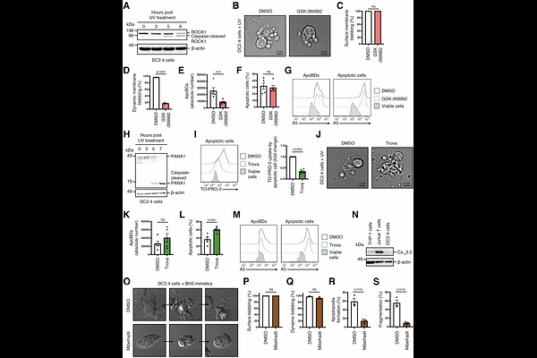
AbstractDendritic cells (DCs) are an important type of antigen presentation cell that regulate immunity by initiating antigen-specific immunity and tolerance through T cell activation. The interaction between DCs and T cells can be mediated through direct cell-cell contact or via the release of extracellular vesicles (EVs) from DCs that harbour antigen presentation machineries. Although small EVs (<200 nm in diameter) such as exosomes released by DCs have been shown to regulate immunity, whether other EV subtypes, in particular those that are released by dying DCs due to homeostatic turnover or following infection, can modulate immune responses is not defined. In this study, we demonstrated that DCs undergoing apoptosis can generate a subclass of large EVs ([~]1,000-5,000 nm in diameter) known as apoptotic bodies (ApoBDs) via distinct morphological steps. Mechanistically, ApoBD formation by apoptotic DCs is regulated by Rho-associated kinase 1 and T-type calcium channels. Functionally, DC-derived ApoBDs were found to mediate direct antigen presentation. These data demonstrate a novel function of ApoBDs and highlight the ability of apoptotic materials derived from dying DCs to continue mediating intercellular communication and regulating immune responses.