Harmonic imaging for nonlinear detection of acoustic biomolecules


Harmonic imaging for nonlinear detection of acoustic biomolecules
Nayak, R.; Duan, M.; Ling, B.; Jin, Z.; Malounda, D.; Shapiro, M. G.
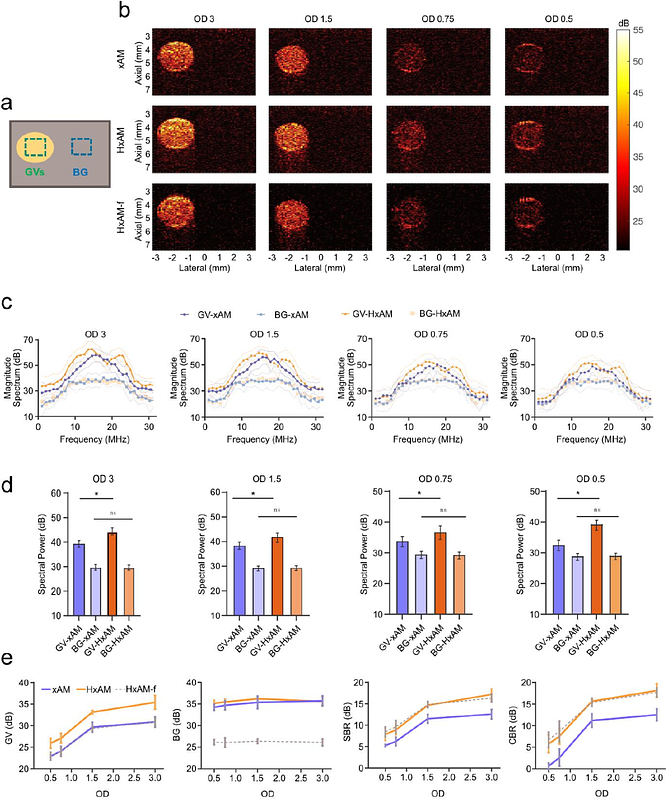
AbstractGas vesicles (GVs) based on acoustic reporter genes have emerged as potent contrast agents for cellular and molecular ultrasound imaging. These air-filled, genetically encoded protein nanostructures can be expressed in a variety of cell types in vivo to visualize cell location and activity or injected systemically to label and monitor tissue function. Distinguishing GVs from tissue signal deep inside intact organisms requires imaging approaches such as amplitude modulation (AM) or collapse-based pulse sequences, however they have limitations in sensitivity or require irreversible collapse of the GVs that restricts its scope for imaging dynamic cellular processes. To address these limitations, this study explores the utility of harmonic imaging to enhance the sensitivity of non-destructive imaging of GVs and cellular processes. Traditional fundamental-frequency imaging utilizing cross-wave AM (xAM) sequences has been deemed optimal for GV imaging. Contrary to this, we hypothesize that harmonic imaging, integrated with xAM could significantly elevate GV detection sensitivity. To verify our hypothesis, we conducted imaging on tissue-mimicking phantoms embedded with purified GVs, mammalian cells genetically modified to express GVs, and live mice after systemic GV infusion. Our findings reveal that harmonic xAM (HxAM) imaging markedly surpasses traditional xAM in isolating GVs\' nonlinear acoustic signature, showcasing significant enhancements in signal-to-background and contrast-to-background ratios across all tested samples. Further investigation into the backscattered spectra elucidates the efficacy of harmonic imaging in conjunction with xAM. HxAM imaging enables the detection of lower concentrations of GVs and cells with ultrasound and extends the imaging depth in vivo by up to 20% and imaging performance metrics by up to 10dB. These advancements bolster the capabilities of ultrasound for molecular and cellular imaging, underscoring the potential of using harmonic signals to amplify GV detection.