Deciphering Coccolith Formation: Advanced Microscopy Insights from the Biomineralisation of Gephyrocapsa huxleyi
Deciphering Coccolith Formation: Advanced Microscopy Insights from the Biomineralisation of Gephyrocapsa huxleyi
Triccas, A.; Verezhak, M.; Ihli, J.; Guizar-Sicairos, M.; Holler, M.; Laidlaw, F.; Singleton, M.; Chamard, V.; Wood, R.; Grunewald, T. A.; Nudelman, F.
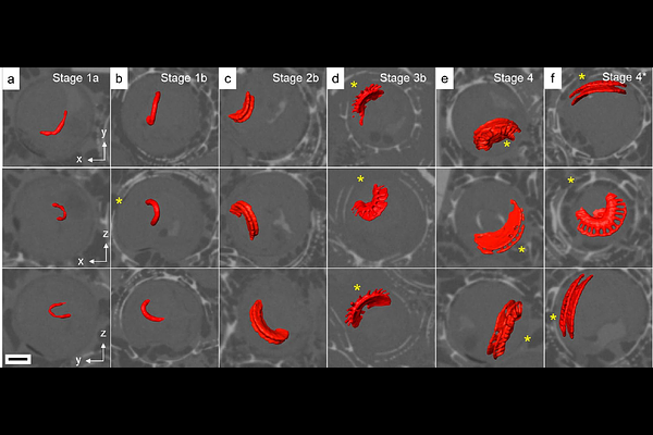
AbstractCoccolithophores are unicellular marine phytoplankton that produce complex and intricately shaped mineralised scales called coccoliths. Coccoliths are produced in an intracellular vesicle where crystal nucleation occurs, from which several individual calcite units develop with anisotropic crystallographic facets, prompting studies into the cellular mechanisms which control crystal growth within the cell. Here, we characterise those morphological developments in 3D that occur during the formation of coccoliths by the species Gephyrocapsa huxleyi using cryo-ptychographic X-ray computed tomography. This technique is ideally suited to study coccolith mineral development, as intracellular structures can be imaged intact in their native state without needing to disrupt cells. Combined with additional imaging of developing coccoliths using cryo-transmission electron microscopy and scanning electron microscopy, we report the developmental stages involved in coccolith growth across the complete mineralisation period, while also showing that the constrained space created by individual crystal units growing in close confinement affects the final crystal morphology and overall mineral structure. These findings provide clarification on the mineralisation pathways that coccolithophores and other biomineralising organisms use to control the formation of highly functionalised crystalline structures, particularly relevant in the design of materials with tunable properties.