Colonization of 3D-organotypic human skin by the Lyme Disease pathogen, Borrelia burgdorferi

Colonization of 3D-organotypic human skin by the Lyme Disease pathogen, Borrelia burgdorferi
Kramer, J. J.; Brinkworth, A.
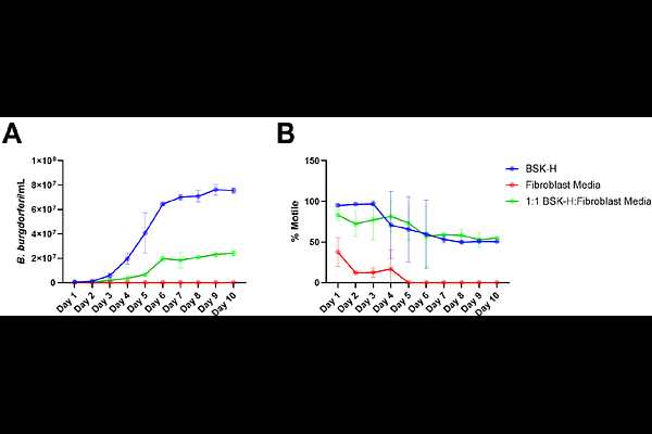
AbstractBorrelia burgdorferi is a bacterial pathogen transmitted by ticks that is the causative agent of Lyme Disease. When studying interactions between B. burgdorferi and skin following a tick-bite, existing models consist primarily of murine skin which has different cellularity and thickness than human skin or ex vivo human biopsies that can be difficult to obtain and can have high variability. This presents a need for a reproducible human skin model. Herein, we adapt an existing human organotypic skin model that is simply composed of dermal fibroblasts and stratified epidermal keratinocytes and develop an infection assay mimicking skin reinfection to characterize B. burgdorferi colonization. Normal spirochete morphology and a stressed B. burgdorferi morphology known as a "round body" were observed. Peak B. burgdorferi invasion was observed at 24 hours (h) with peak round body formation at 48 h. By breaking the skin down into its individual components, we observed an increase in the number of round bodies in the presence of dermal fibroblasts. The presence of keratinocytes or extracellular matrix alone had no effect on round body formation, indicating a dermal fibroblast-mediated mechanism. We also demonstrate tissue-to-tissue dissemination and colonization, setting the groundwork for future studies with other tissues. Collectively, these results prove that this is a valid human skin model to study B. burgdorferi colonization during secondary dissemination.