Spatial transcriptomics from pancreas and local draining lymph node tissue reveals a lymphotoxin-β signature in human type 1 diabetes
Spatial transcriptomics from pancreas and local draining lymph node tissue reveals a lymphotoxin-β signature in human type 1 diabetes
Medina-Serpas, M. A.; Brusko, M.; Golden, G. J.; Campbell-Thompson, M.; Rogers, T.; Posgai, A. L.; Luning-Prak, E. T.; Kaestner, K. H.; Betts, M. R.; McIntyre, L. M.; Atkinson, M. A.; Brusko, T. M.
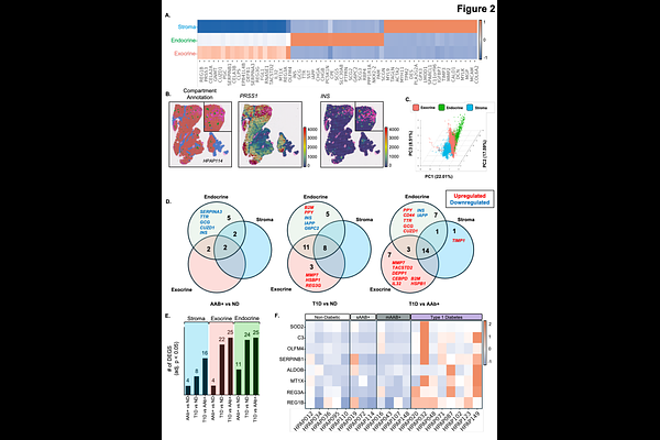
AbstractThis study explores the inflammatory response observed in pancreata and pancreatic lymph node (pLN) samples obtained throughout the natural history of type 1 diabetes (T1D) including non-diabetic individuals and non-diabetic autoantibody positive individuals with high susceptibility using spatial transcriptomics (ST). Integration of ST with public single-cell RNA sequencing data enabled interrogation of transcriptional alterations in T1D pathogenesis across both tissues and cellular scales. In the T1D pancreas, we observed global upregulation of multiple inflammation-associated transcripts, including regenerating islet-derived (REG) family genes, complement factor 3 (C3), SOD2, and OLFM4, and highlighted cellular candidates potentially contributing to these signatures. Within the T1D pLN, we observed spatially restricted upregulation of lymphotoxin-{beta} (LTB) alongside follicular dendritic cell (FDC)-associated transcripts including FDCSP, CLU, and FCER2. Collectively, these findings highlight a distinct inflammation signatures in the pancreas and regional pLN which can help inform the development of future therapeutic interventions.