Micro-elastography of biopsies
Micro-elastography of biopsies
Gregoire, S.; Giammarinaro, B.; Le Quere, D.; Devissi, M.; BRULPORT, A.; Catheline, S.
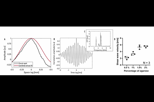
AbstractMicro-elastography is an optical technique that studies elastic waves for the mechanical characterisation of micrometric objects, such as cells. We propose to adapt this technique for the characterisation of millimetre-sized samples using a white light microscope. The objective is to perform a rapid, global characterisation of the elasticity of a biopsy. The millimetre-sized samples to be characterized are embedded in an agarose gel. A vibrator generates shear waves in this gel that transmit naturally inside the sample. This technique removes the need for precise manipulation of the wave source. A high-speed camera records the propagation of the waves in the sample. Their velocity is calculated using a noise correlation approach. Due to the lack of millimetric phantoms of calibrated elasticity, we choose to validate this method with a three step process. The experimental setup is first validated on homogeneous gels, then on biological samples of increasing elasticity, biopsies of beef liver hardened by heating, and finally on biological samples of clinical interest: biopsies of mouse endometrium. This method can be applied to all types of biological tissue, paving the way for rapid mechanical characterization of biopsies.