Metabolic Analysis of Human Retinal Pigment Epithelium and Choroid Tissue in Aging and Macular Degeneration
Metabolic Analysis of Human Retinal Pigment Epithelium and Choroid Tissue in Aging and Macular Degeneration
Navratil, E. M.; Liu, X.; Wiley, L. A.; Anderson, M. G.; Meyer, K. J.; Brown, R. F.; Evans, I. A.; Taylor, E. B.; Stone, E. M.; Tucker, B. A.; Mullins, R. F.
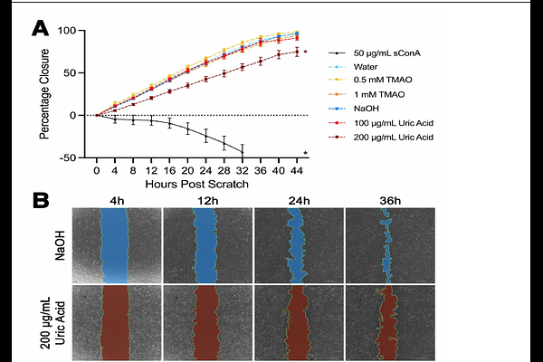
AbstractAge-related macular degeneration is a common ocular disease that causes vision loss in the elderly, with a complex set of risk factors and proposed mechanisms of pathogenesis. A powerful method for investigating changes in disease is metabolomics, by which small molecules can be identified and quantified simultaneously. We report here the metabolic analysis of human RPE-choroid tissue in aging and macular degeneration (AMD), as well as comparisons of human macular and extramacular RPE-choroid and neural retina. Levels of 215 metabolites were determined in young donors, AMD donors (early/intermediate, geographic atrophy, and neovascularization) and age-matched controls. The largest number of metabolite differences were observed between young and healthy aged controls, as opposed to between aged controls and any stage of AMD. Two notable metabolites found to be increased in aging choroids are trimethylamine N-oxide and uric acid, both of which were significant after Bonferroni correction. A mouse endothelial cell line treated with a high concentration of uric acid exhibited reduced migration in a wound closure assay. This study provides initial insights into the metabolome of human choroids in varying states of age and macular degeneration, as well as functional implications of these changes in the aging choroid.