Semi-automated reconstruction of glomerular architecture from 3D confocal microscopy data

Semi-automated reconstruction of glomerular architecture from 3D confocal microscopy data
Loyd, Y. M.; Chase, S. E.; Krendel, M.
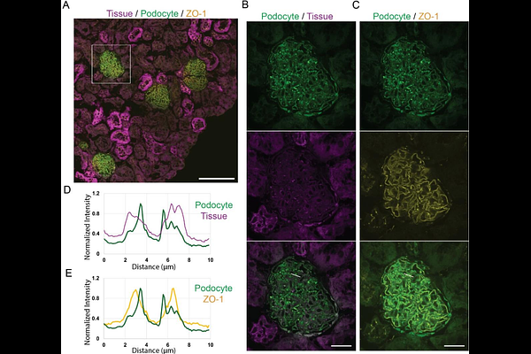
AbstractNephrons are the functional units of the kidney; within each nephron, the glomerulus is the initial site of selective filtration that allows removal of waste products while preserving proteins in the bloodstream. Each glomerulus consists of a network of capillaries surrounded by specialized epithelial cells, podocytes, which mediate selective filtration. Abnormalities in glomerular structure impair renal function, resulting in proteinuria and kidney disease. Although several microscopy-based approaches exist to characterize glomerular architecture and structural abnormalities, quantitative analysis is often limited by labor-intensive image segmentation. In this study we present a semi-automated approach for segmentation and analysis of glomerular architecture from three-dimensional confocal microscopy data. Using mTmG transgenic mice that express membrane-associated EGFP in podocytes and membrane-associated tdTomato across all other cell types, we reconstruct podocyte processes and glomerular capillaries from volumetric renal images. This semi-automated approach reduces manual segmentation effort and supports more efficient, standardized analysis of glomerular architecture in three-dimensional confocal microscopy datasets.