Tunable 3D Alveolosphere Model from Human Alveolar Cells: A Breakthrough Tool to Explore Emphysema Pathophysiology
Tunable 3D Alveolosphere Model from Human Alveolar Cells: A Breakthrough Tool to Explore Emphysema Pathophysiology
Guecamburu, M.; Pavot, A.; Jeanniere, c.; belaroussi, y.; Thumerel, M.; sammaniego, e.; begueret, H.; maucort, g.; decoeur, f.; Dupuy, J.-W.; raymond, a.-a.; esteves, P.; Grassion, L.; dournes, G.; berger, p.; maurat, e.; latouille, e.; Studer, V.; Dupin, I.; henrot, P.; zysman, m.
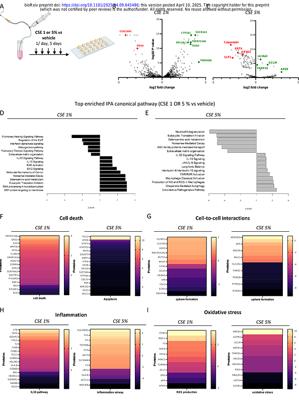
AbstractRationale: Three-dimensional (3D) culture models such as alveolosphere, provide a unique tool to study emphysema mechanisms. Reducing the heterogeneity of alveolospheres which are currently mostly grown in Matrigel remains challenging. Objectives: To develop a tunable and reproducible 3D-alveolosphere model exclusively from human primary type II alveolar epithelial cells (AEC2) for modeling and understanding emphysema. Methods: AEC2 cells (HTII-280+) were isolated from 52 smoker and non-smoker lung samples and cultured in preformed photopolymerized hydrogel microwells of adjustable shape and size. Topological and phenotypic characterization were performed at Day (D)1, 7 and 14. Lamellar bodies (LB) were quantified using artificial intelligence (AI)-based image analysis of transmission electron microscopy (TEM) serial block face images. Emphysema was modeled through chronic exposure to 1 and 5% cigarette smoke extract (CSE) during 5 consecutive days. Measurements and Main Results: 3D-alveolospheres were maintained in culture for 14 days with central lumen formation observed from D7 to D14. Presence of tight junctions (TEM imaging and ZO-1 immunostaining) suggested epithelial barrier formation. AEC1 markers (p2xr4, pdpn) appeared progressively from D1 to D14 while AEC2 markers (abca3, sftpa, sftpc) persisted over time. TEM images indicated surfactant synthesis (LB, lipid bodies) and AI-driven LB quantification showed a decrease in the proportion of LB-containing cells over time. CSE exposure led to cell death, architectural disorganization, oxidative stress and inflammation. Conclusion: This standardized and adjustable 3D-alveolosphere model from human primary AEC2, reproduced key native alveolar features. CSE exposure provides an opportunity to appropriately study the pathophysiological pathways involved in emphysema.