Anatomical and Molecular Characterization of the Zebrafish Meninges

Anatomical and Molecular Characterization of the Zebrafish Meninges
Venero Galanternik, M.; Castranova, D.; Gober, R. D.; Nguyen, T.; Kenton, M.; Margolin, G.; Kraus, A.; Sur, A.; Dye, L. E.; Pham, V.; Maese, A.; Holmgren, M.; Gore, A. V.; Samasa, B.; Goldstein, A.; Davis, A. E.; Swearer, A. A.; Iben, J. R.; Li, T.; Coon, S. L.; Dale, R. K.; Farrell, J. A.; Weinstein, B. M.
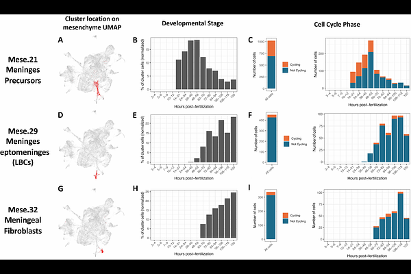
AbstractThe meninges are a set of connective tissue layers that surround the central nervous system, protecting the brain from mechanical shock, supporting its buoyancy, guarding it from infection and injury, and maintaining brain homeostasis. Despite their critical role, the molecular identity, developmental origins, and functional properties of the cell types populating the meninges remain poorly characterized. This is in large part due to lack of cell type specific markers and difficulty in visualizing and studying these structures through the thick mammalian skull. Here, we show that the zebrafish, a genetically and experimentally accessible vertebrate, possesses an easily imaged mammalian-like meninges. Anatomical and cellular characterization of its composition via histology, electron microscopy, and confocal imaging shows that the adult zebrafish possesses complex multilayered meninges with double-layered dura mater and intricate leptomeningeal layers. Using single cell transcriptomics, we define the molecular identities of meningeal cell populations, including a unique ependymin (epd)-expressing cell population that constitutes the major cellular component of the leptomeningeal barrier and is essential for brain development and survival. These findings support the use of zebrafish as a useful comparative model for studying the meninges, provide a foundational description for future zebrafish meningeal research, and identify a new Leptomeningeal Barrier Cell that serves as the primary epithelial cell component of the leptomeninges.