Evidence for reduced choroid plexus volume in the aged brain
Evidence for reduced choroid plexus volume in the aged brain
Youh, R.-Y.; Perera, C.; Harrison, I. F.; Lythgoe, M. F.; Wright, D. K.; Nizari, S.; Wells, J. A.
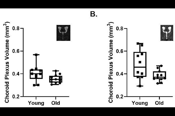
AbstractBackground: The choroid plexus plays an important role in brain homeostasis, including the active secretion of cerebrospinal fluid. Its function and structure have been reported to be affected by normal ageing. However, existing measures of choroid plexus volume may be complicated by partial volume (in-vivo MRI) and tissue fixation artefacts (histology). In this study, we investigate possible changes in choroid plexus volume within the lateral ventricles of a mouse model of ageing utilising two structural MRI protocols explicitly designed for time-efficient, high-resolution in-vivo imaging of the choroid plexus. Methods: Two MRI sequences were utilised to examine in-vivo choroid plexus volume in the lateral ventricles of young (~6 months) and aged (~24 months) mouse brains: 1) an ultra-long echo-time T2 weighted fast-spin-echo and 2) a multi-TE T2* mapping protocol. A test-retest study was performed on a subset of the data to examine the reproducibility of choroid plexus volume estimation. A two-way ANOVA test was performed to determine possible differences in choroid plexus volume in young and aged mouse groups across the two distinct MRI protocols. Results: Reproducibility tests showed a low test-retest variability of the manual segmentation pipeline for both MRI protocols. A statistically significant reduction of in vivo choroid plexus volume was found in the aged mouse brain. This finding is concordant with previous histology studies that have observed a reduction in epithelial cell height with ageing across a wide range of species. Conclusions: Here, we present an in vivo investigation of changes to lateral ventricle choroid plexus volume in the mouse brain utilising a manual segmentation approach based on two bespoke MRI protocols designed for time-efficient high resolution imaging of the choroid plexus. We provide evidence for a reduction in choroid plexus volume in the aged brain. This research provides insight for studies utilising MRI measurements of choroid plexus volume as a biomarker of age-related neurologic conditions as it indicates that the ageing process itself does not result in hypertrophy of the choroid plexus.