Planar Amorphous Silicon Carbide Microelectrode Arrays for Chronic Recording in Rat Motor Cortex
Planar Amorphous Silicon Carbide Microelectrode Arrays for Chronic Recording in Rat Motor Cortex
Abbott, J. R.; Jeakle, E. N.; Haghihi, P.; Usoro, J. O.; Sturgill, B. S.; Wu, Y.; Geramifard, N.; Radhakrishna, R.; Patnaik, S.; Nakajima, S.; Hess, J.; Mehmood, Y.; Devata, V.; Vijayakumar, G.; Sood, A.; Thai, T. T. D.; Dogra, K.; Hernandez-Reynoso, A. G.; Pancrazio, J. J.; Cogan, S. F.
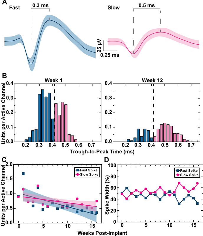
AbstractChronic implantation of intracortical microelectrode arrays (MEAs) capable of recording from individual neurons can be used for the development of brain-machine interfaces. However, these devices show reduced recording capabilities under chronic conditions due, at least in part, to the brain\'s foreign body response. This creates a need for MEAs that can minimize the foreign body response to enable long-term recording. A potential approach to reduce the foreign body response is the use of ultrathin MEAs. Here, we fabricated ultrathin (cross-sectional area: 160 um2) amorphous silicon carbide (a-SiC) MEAs with sixteen electrode channels and implanted them into the motor cortex of seven female Sprague-Dawley rats. A-SiC was chosen as the fabrication base for its high chemical stability, good electrical insulation properties, and amenability to thin film fabrication techniques. Electrochemical analysis and neural recordings were performed weekly for 4 months. MEAs were characterized in vitro pre-implantation and in vivo using electrochemical impedance spectroscopy and cyclic voltammetry at 50 mV/s and 50,000 mV/s. Neural recordings were analyzed for single unit activity. At the end of the study, animals were sacrificed for immunohistochemistry analysis. We observed statistically significant, but small, increases in 1 and 30 kHz impedance values and 50,000 mV/s charge storage capacity over the 16-week implantation period. Slow sweep 50 mV/s CV and 1 Hz impedance did not significantly change over time. Impedance values increased from 11.6 MOhms; at 30 kHz over 16 weeks. The median charge storage capacity of the implanted electrodes at 50 mV/s were 58.1 mC/cm2 on week 1 and 55.9 mC/cm2 on week 16, and at 50,000 mV/s were 4.27 mC/cm2 on week 1 and 5.93 mC/cm2 on week 16. Devices were able to record neural activity from 92% of all active channels at the beginning of the study, At the study endpoint, a-SiC devices were still recording single-unit activity on 51% of electrochemically active electrode channels. In addition, we observed that the signal-to-noise ratio experienced a small decline of only -0.19 per week. We also classified the units as fast and slow spiking based on the trough-to-peak time. Although the overall presence of single units declined, fast and slow spiking units declined at a similar rate. Furthermore, immunohistochemistry showed minimal foreign body response to the a-SiC devices, as highlighted by statistically insignificant differences in activated glial cells between implanted brains slices and contralateral sham slices, as evidenced by GFAP staining. NeuN staining revealed the presence of neural cell bodies close to the implantation site, again statistically not different from a contralateral sham slice. These results support the use of ultrathin a-SiC MEAs for long-term implantation and use in brain-machine interfaces.