High precision fluorescence tomography-guided system for pre-clinical radiation research: system design and validation
High precision fluorescence tomography-guided system for pre-clinical radiation research: system design and validation
Xu, X.; Tseng, Y.; Deghhani, H.; Hardy, L.; Tong, Z.; Guo, G.; Habib, A.; Iordachita, I.; Wong, J.; Wang, K. K.-H.
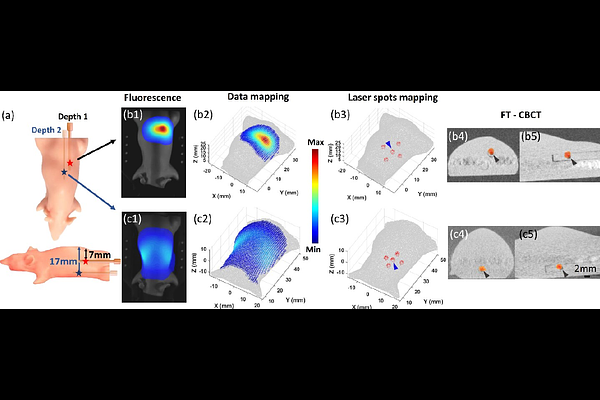
AbstractPurpose: Cone-beam computed tomography (CBCT), commonly used for image guidance in pre-clinical studies, is limited in soft tissue localization and lacks the functional information needed for image-guided radiation studies and treatment assessment. To address these limitations, we developed 3D fluorescence tomography (FT) for high precision functional image-guided research and integrated it with small animal irradiators. Methods: The FT system, integrated with a commercial bioluminescence-guided platform in a standalone configuration, enabling compact multi-projection and multi-spectral imaging. A dual-axis galvo mirror scanner was used for laser spots scanning to excite internal fluorophore. A transportable mouse bed allows animals imaged in the optical system and transferred to irradiators for CBCT imaging and FT-guided irradiation. Spatial geometry-based methods were developed to map the laser spots and fluorescence images onto the animal numerical mesh surface, generated from the CBCT image, to serve as input data for FT reconstruction. A self-calibration method using Born-ratio data, fluorescence normalized to the excitation image, was adopted for reconstruction. Mouse phantom embedded with QD800 reporter and orthotopic glioblastoma (GBM) model injected with IRDye 800CW labelled U87-EGFR cells were used to verify the FT-guided system in target localization. Results: The laser spot mapping accuracy are within 0.7 mm maximum deviation. Our system can reconstruct the target QD800 as deep as 17 mm in the mouse phantom with single projection fluorescence imaging at localization accuracy < 1 mm deviation. Using the Born ratio approach, we effectively eliminated excitation light leakage and autofluorescence, enabling our FT system to localize IRDye 800CW-labeled EGFR-overexpressing GBM cells in the mouse brain with approximately 1 mm accuracy. Conclusion: We developed a compact FT system integrated with a small animal irradiator, achieving sub-millimeter localization accuracy in phantom and in vivo models. Its precision, flexibility, and compatibility position it as a powerful tool for advancing functional imaging-guided pre-clinical radiation research.