Optimizing effective labeling efficiency in MINFLUX 3D DNA-PAINT microscopy by maximizing marker detection probability.
Optimizing effective labeling efficiency in MINFLUX 3D DNA-PAINT microscopy by maximizing marker detection probability.
Soeller, C.; Bokhobza, A. F. E.; Casares-Arias, J.; Clowsley, A. H.
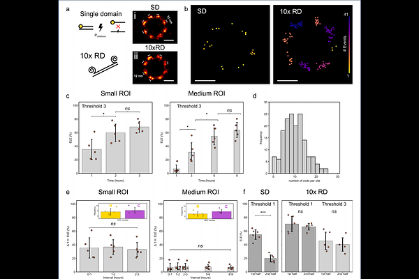
AbstractMINFLUX is a powerful single-molecule approach capable of achieving high spatially isotropic resolution in three dimensions. Current implementations collect localizations strictly serially, but criteria for when to terminate acquisition are often unclear. We therefore systematically investigate the time course of effective labeling efficiency (ELE) and achievable saturation values in MINFLUX 3D DNA-PAINT microscopy of Nup96 proteins in a U-2 OS-Nup96-mEGFP modified cell line using a commercial MINFLUX microscope. ELE was measured with a maximum-likelihood template fitting assisted quantitative procedure. We collected data measured over various scan sizes and achieved ELE values of ~60% after passing a time interval dependent on the region size, typically requiring long-duration acquisitions over several hours. Our data and a simple model suggest that maximizing marker detection is key to achieving the limits set by chemical labeling efficiency. A factor limiting the marker detection probability when using conventional DNA-PAINT markers is docking strand site-loss, observed over the duration required to build up the image data of MINFLUX acquisitions, which also limits the achievable number of labeling site visits to values around 1-3. Using repeat DNA-PAINT, i.e. employing oligonucleotide sequences with repeated docking sites, we observed greatly reduced site-loss and could increase the number of individual visits to site locations by more than threefold over the same period. Additionally, this enabled increasing stringency criteria for labeling (i.e. higher threshold values) and maximizing marker detection probabilities so that ELE reaches the limits set by chemical labeling efficiency.