Boldine prevents diabetes-induced skeletal muscle dysfunction by inhibiting large-pore channels
Boldine prevents diabetes-induced skeletal muscle dysfunction by inhibiting large-pore channels
VASQUEZ, W.; Cea, L. A.; Troncoso, F.; Sandoval, H.; Lira, A.; Figueroa, X.; Escudero, C.; Saez, J. C.
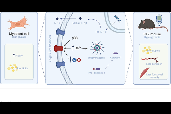
AbstractBackground Diabetes mellitus leads to skeletal muscle dysfunction associated with loss of strength, impaired blood perfusion, lipid accumulation, and inflammation. The opening of large-pore channels has been linked to increased membrane permeability and inflammatory signaling in several pathologies. Boldine, an alkaloid from Peumus boldus, blocks large-pore channel activity and exhibits antioxidant and anti-inflammatory properties. This study evaluated whether boldine prevents skeletal muscle alterations induced by diabetes and explored potential underlying mechanisms. Methods Diabetes was induced in male C57BL/6J mice using streptozotocin (STZ, 40 mg/kg/day for 5 days). Diabetic mice were treated with boldine (50 mg/kg/day) for four weeks. Muscle strength and resting membrane potential were analyzed in vivo. Also, right gastrocnemius muscle blood perfusion at basal and after acetylcholine (10 M) stimulation were analyzed in vivo. Lipid accumulation was assessed using Oil Red O staining, and CD31 immunodetection was used to evaluate capillary density. mRNA levels of NLRP3 were evaluated in muscle by qPCR. In human myoblasts (AB1167) cultured under low (8 mM) or high glucose (25 mM) conditions, with or without boldine, membrane permeability (ethidium uptake), intracellular Ca2+ (Fura-2), nitric oxide (DAF-FM), and levels of NLRP3 and Casp1 (qPCR) and reactivity PPAR gamma; (Immunofluorescence) were determined. Results STZ mice showed reduced muscle strength and depolarized resting membrane potential, both prevented by boldine. Basal muscle perfusion was ~20% lower in diabetic mice (160.1 {+/-} 17.2 vs. 199.1 {+/-} 13.8 units), whereas boldine preserved perfusion (184.6 {+/-} 14.3 units). Oil Red O-positive fibers increased to 52.4 {+/-} 3.6% in diabetic mice and decreased to 15.2 {+/-} 4.1% with boldine (control: 3.1 {+/-} 1.3%; p<0.05). NLRP3 mRNA increased 17.7 {+/-} 2.8-fold in diabetic muscle and was reduced by ~50% with boldine. In myoblasts, high glucose increased ethidium uptake, nitric oxide production, NLRP3 and caspase-1 expression, and nuclear PPAR gamma (~45% positive nuclei); all effects were prevented by boldine. Conclusions Boldine preserves skeletal muscle function and vascular reactivity in diabetes and prevents lipid accumulation and inflammasome activation both in vivo and in vitro. These effects are associated with inhibition of large-pore channel activity and attenuation of downstream calcium-dependent, inflammatory, and adipogenic pathways, supporting boldine as a promising therapeutic candidate for diabetes-associated skeletal muscle dysfunction.